Search & Filter All Posts
All Articles
(6 Results)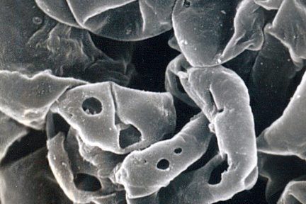
Basement Membrane Damage in Crescentic GN
Todays eyeSCANdy image shows acellular scanning EM showing GBMs with variably sized discrete perforations. This is the mildest form of…
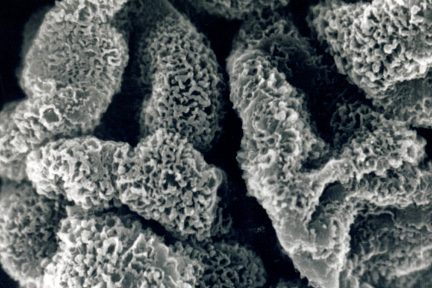
Membranous Glomerulonephritis Stage II
This eyeSCANdy image shows acellular scanning EM from a biopsy with membranous glomerulonephritis, stage II. The extracted deposits previously resided…
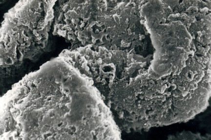
Stage III Membranous Glomerulonephritis
Todays eyeSCANdy shows acellular scanning EM from a biopsy with membranous glomerulonephritis, stage III! There is more extensive GBM bridging…
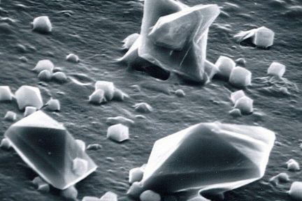
Oxalate Crystals In A Urinary Sediment
This eyeSCANdy image shows calcium oxalate crystals in a urinary sediment under scanning electron microscopy! Image courtesy of Dr.…
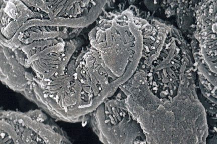
Glomerular Capillary Loops with Interdigitating Podocyte Foot Processes
This eyeSCANdy image shows scanning EM of normal glomerulus showing glomerular capillary loops with interdigitating podocyte foot processes! Image courtesy…
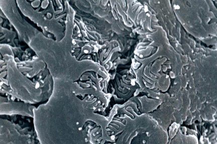
Scanning EM of a Proteinuric Glomerulus
This eyeSCANdy image shows scanning EM of a proteinuric glomerulus with capillary loop showing widening and partial loss of interdigitating…