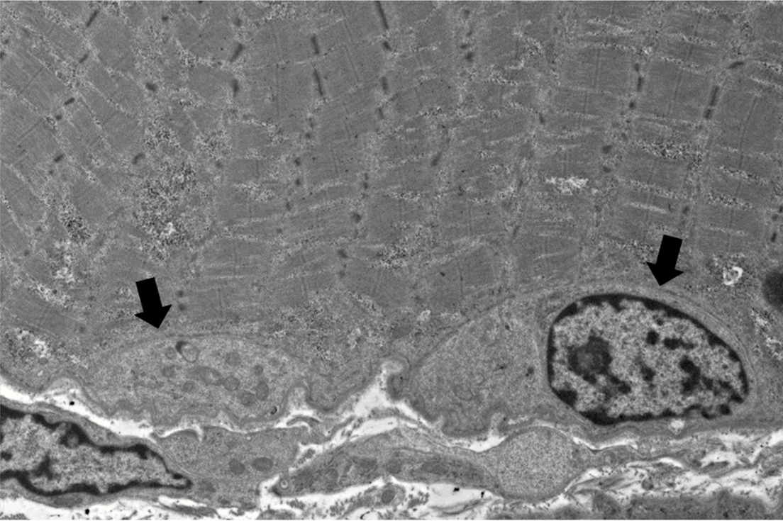
An adult patient with proximal muscle weakness presents with CK 4400 and skeletal muscle biopsy of the thigh shows frequent regenerating and necrotic muscle fibers. In this electron micrograph of skeletal muscle, what are the structures indicated by the black arrows?
Satellite cells are a normal population of undifferentiated cells within a skeletal muscle fiber that is capable of differentiating into myoblasts (i.e. stem cell-like properties), hence providing a source to produce new myotubes, as well as regeneration after injury (i.e. necrotizing myopathy). The figure demonstrates satellite cells between the basal lamina and plasma membrane. One of the satellite cells contains a nucleus with heterochromatic chromatin, and its cytoplasm contains organelles (i.e. mitochondria and lysosomes). See references.
References:
Dubowitz V, Sewry CA, Oldfors A. Normal Muscle. In: Muscle Biopsy: A Practical Approach, 5th ed. Saunders Elsevier, London, United Kingdom; 2021: 35-37.
Yin H, Price F, Rudnicki MA. Satellite cells and the muscle stem cell niche. Physiol Rev. 2013 Jan;93(1):23-67. PMID: 23303905.
Quick note: This post is to be used for informational purposes only and does not constitute medical or health advice. Each person should consult their own doctor with respect to matters referenced. Arkana Laboratories assumes no liability for actions taken in reliance upon the information contained herein.