Clinical History:
Neuropathologic examination (brain autopsy) on this 65-year-old patient with a history of slowly progressive dementia demonstrated gross and microscopic findings consistent with Alzheimer’s disease.
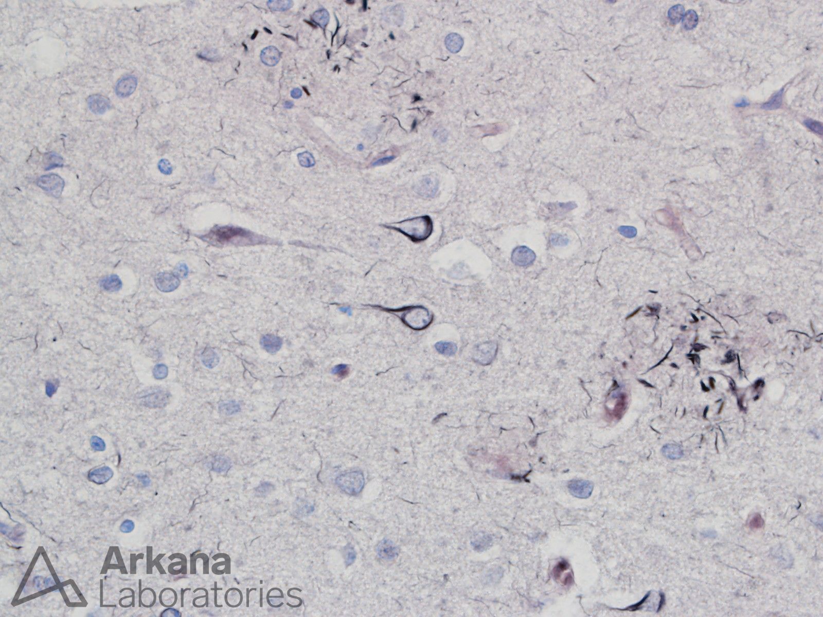
Can you name the three types of paired helical filament accumulations (Tau protein) demonstrated on the provided Gallyas silver stain image?
Answer:
The Gallyas preparation nicely highlights flame shaped neurofibrillary tangles, variable thick bands within Abeta amyloid containing neuritic plaques, and numerous background fine neuropil threads.
Reference(s) / Additional Reading:
- Braak H, Braak E, Grundke-Iqbal I, Iqbal K. Occurrence of neuropil threads in the senile human brain and in Alzheimer’s disease: a third location of paired helical filaments outside of neurofibrillary tangles and neuritic plaques. Neurosci Lett. 1986 Apr 24;65(3):351-5. doi: 10.1016/0304-3940(86)90288-0. PMID: 2423928.
- Kuninaka N, Kawaguchi M, Ogawa M, Sato A, Arima K, Murayama S, Saito Y. Simplification of the modified Gallyas method. Neuropathology. 2015 Feb;35(1):10-5. doi: 10.1111/neup.12144. Epub 2014 Sep 1. PMID: 25178396; PMCID: PMC4491351.
Quick note: This post is to be used for informational purposes only and does not constitute medical or health advice. Each person should consult their own doctor with respect to matters referenced. Arkana Laboratories assumes no liability for actions taken in reliance upon the information contained herein.