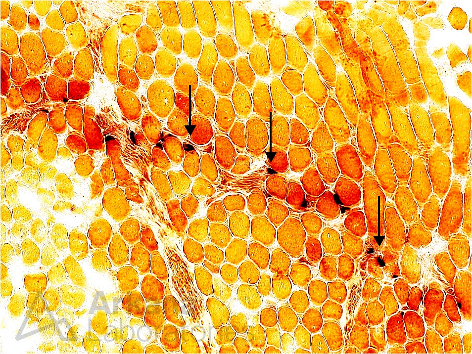
Neuromuscular junctions in normal skeletal muscle. Esterase preparation (A) darkly highlights neuromuscular junctions (NMJs) along a small intramuscular nerve twig. Original magnification: A. Nonspecific Esterase Preparation, 100x.
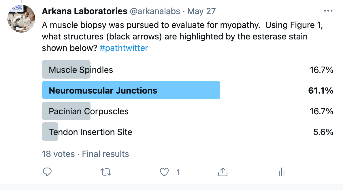
A relatively young patient presented with hypotonia. Course features were noted on physical exam; however, multiple studies revealed no etiology for the patient’s clinical picture. A muscle biopsy was pursued to evaluate for myopathy. Using Figure 1, what structures (black arrows) are highlighted by the esterase stain shown below?
Answer: Neuromuscular Junctions
Nonspecific esterase preparation is one of the principal enzymatic histochemical stains used to evaluate neurogenic pathology in skeletal muscle by way of darkly highlighting acutely angulated atrophic muscle fibers with over-reactivity of staining signifying denervation-type change that is active/ongoing.
In this case, esterase preparation darkly highlights neuromuscular junctions (NMJs) along a small nerve twig. Evaluation for the presence of these normal structures may prompt additional study for nerve twigs on the plastic embedded thick sections, and subsequent search to evaluate the NMJ by electron microscopy.
Neuromuscular junctions ultrastructural evaluation may reveal abnormalities of the organization of the apparatus that could be helpful in determining the etiology of neuromuscular disorders (i.e. loss of postsynaptic complexity in myasthenia gravis).
Why were the other answers wrong?
The other answer choices in this case are also normal intramuscular structures that could be randomly/incidentally found in a skeletal muscle biopsy. It is important for the neuropathologist to recognize these normal structures, as to not be lead down an incorrect diagnostic odyssey.
Muscle spindles are part of the body’s reflex apparatus.
Likewise, Pacinian corpuscles are part of the body’s sensory system to pressure and vibration.
Myotendinous insertion sites may also darkly highlight with nonspecific esterase, but these are found near dense fibrous connective tissue anchoring the muscle fiber. NMJs are usually found adjacent to and nearby small nerve twigs.
References
Cai C, Anthony DC, Pytel P. A pattern-based approach to the interpretation of skeletal muscle biopsies. Mod Pathol. 2019 Apr;32(4):462-483. PMID: 30401945.
Conti-Fine BM, Milani M, Kaminski HJ. Myasthenia gravis: past, present, and future. J Clin Invest. 2006 Nov;116(11):2843-54. PMID: 17080188.
Dubowitz V, Sewry CA, Oldfors A. Muscle Biopsy: A Practical Approach, 5th ed. Saunders Elsevier, London, United Kingdom; 2021.
Nix JS, Moore SA. What Every Neuropathologist Needs to Know: The Muscle Biopsy. J Neuropathol Exp Neurol. 2020 Jul 1;79(7):719-733. PMID: 32529201.
Quick note: This post is to be used for informational purposes only and does not constitute medical or health advice. Each person should consult their own doctor with respect to matters referenced. Arkana Laboratories assumes no liability for actions taken in reliance upon the information contained herein.