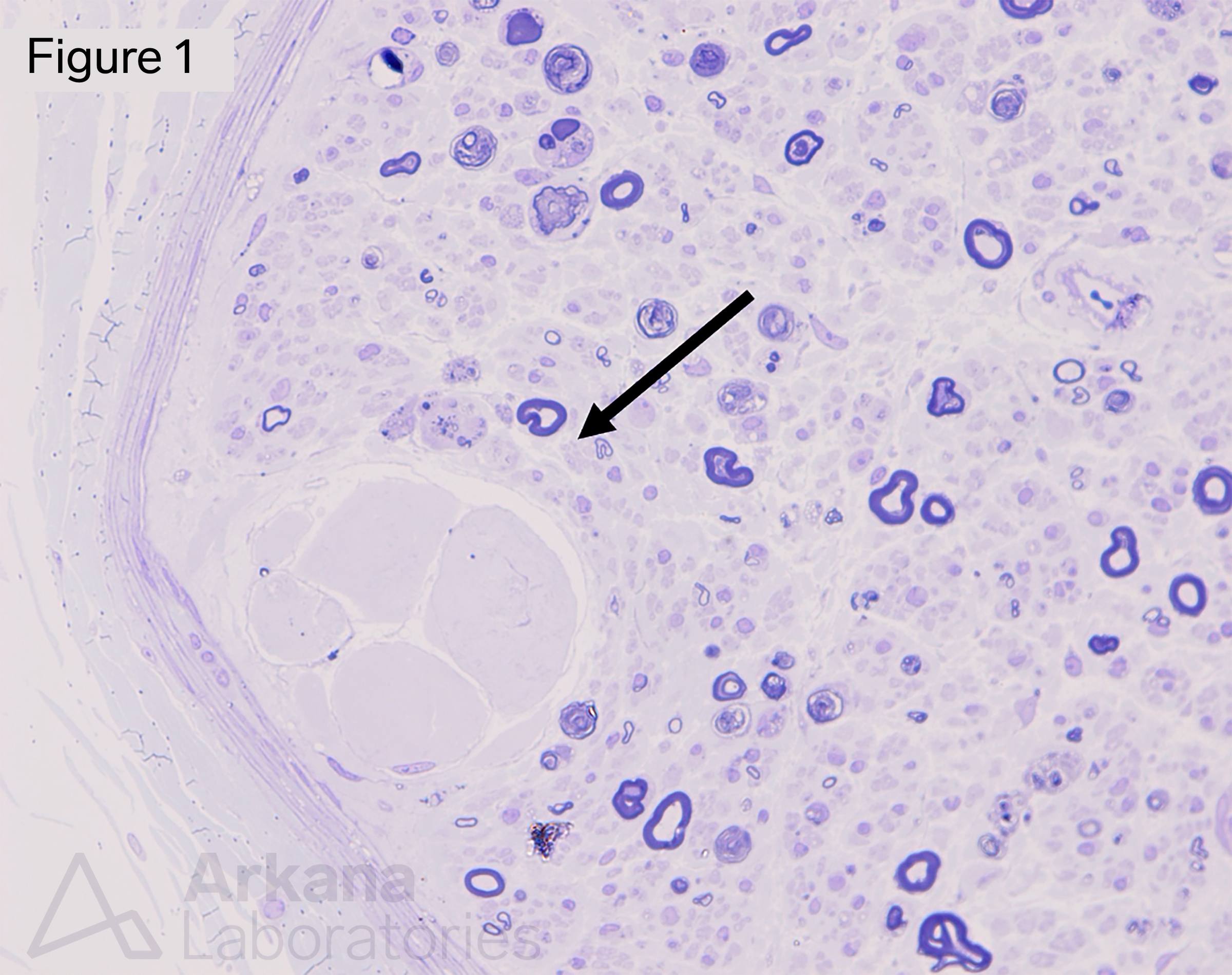
Figure 1: Cross section of sural nerve with Renaut Body (arrow) and background moderate diffuse axonopathic process. Plastic embedded thick section stained with Toluidine blue, original magnification x400.
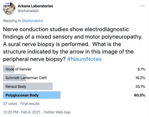
A 65-year-old female presents with complaint of numbness and tingling in her hands and feet with gradual progressive onset. She is diabetic, and clinical work-up shows no positive autoantibodies. Nerve conduction studies show electrodiagnostic findings of a mixed sensory and motor polyneuropathy. The clinical differential diagnosis includes diabetic polyneuropathy versus chronic inflammatory demyelinating polyradiculoneuropathy (CIDP). A sural nerve biopsy is performed. What is the structure indicated by the arrow in this image of the peripheral nerve biopsy?
Answer: The indicated structure is a Renaut Body. In peripheral nerve biopsies, this benign normal structure is seen in about 2% to 7% of specimens, and familiarity with this common finding is important in order to avoid diagnostic errors. Renaut bodies are round to ovoid, whorled structures, predominantly located in the subperineurial region. Renaut bodies do not contain axons, and their most common cellular constituent is the fibroblast. The literature has suggested a variable association with formation in response to mechanical stress and hypothyroid neuropathy; however, studies are still ongoing. See references.
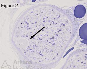
Figure 2: Cross section of sural nerve with Renaut Body (arrow) and background moderate diffuse axonopathic process. Plastic embedded thick section stained with Toluidine blue, original magnification x200.
Why were the other answers incorrect?
The node of Ranvier is a myelin sheath gap that is part of the micro-neuroanatomy facilitating saltatory conduction, best seen in longitudinal section by electron microscopy.
Schmidt-Lanterman incisures or clefts are transverse regions of uncompact myelin by Schwann cell cytoplasm that are believed to serve as a mechanism for inner and outer cellular communication, best seen in longitudinal section by electron microscopy.
Polyglucosan bodies (PGBs) variably sized cytoplasmic inclusions that appear basophilic with H&E stain by routine light microscopy and are somewhat metachromatic with Toluidine blue. In near-normal nerve biopsies, one or two PGBs may be seen in approximately 15% of specimens, especially in nerve biopsies from more elderly patients.
References:
Averback P, Langevin H. Corpora amylacea of the lumbar spinal cord and peripheral nervous system. Arch Neurol. 1978;35: 95-96.
Bergouignan FX, Vital C. Occurrence of Renaut’s bodies in a peripheral nerve. Arch Pathol Lab Med. 1984 Apr;108(4):330-3. PMID: 6546675.
Bilbao JM and Schmidt RE. Normal anatomy of the peripheral (sural) nerve. Biopsy diagnosis of peripheral neuropathy, 2nd ed. Springer, Switzerland. 2015: 21-42.
Busard, H.L.S.M., Gabreëls-Festen, A.A.W.M., van ‘t Hof, M.A. et al. Polyglucosan bodies in sural nerve biopsies. Acta Neuropathol. 1990;80: 554-557.
Ghabriel MN and Allt G. Incisures of Schmidt-Lanterman. Progress in Neurobiology. 1981;17: 25-58.
MacKinnon SE, Dellon AL, Hudson AR, Hunter DA. Chronic human nerve compression – a histological assessment. Neuropathology and Applied Neurobiology. 1986;12: 547-565.
Quick note: This post is to be used for informational purposes only and does not constitute medical or health advice. Each person should consult their own doctor with respect to matters referenced. Arkana Laboratories assumes no liability for actions taken in reliance upon the information contained herein.