This 50-year old immunocompromised patient died after presenting with rapid cognitive decline and signs of brainstem dysfunction (cranial nerve deficits). MRI demonstrated multiple ring-enhancing cerebral and brainstem lesions. One of the lesions was biopsied.
What is you diagnosis based on Figures #1 to #3?
A. Toxoplasmosis
B. Acanthamoeba
C. HSV1/2
D. Coccidioidomycosis
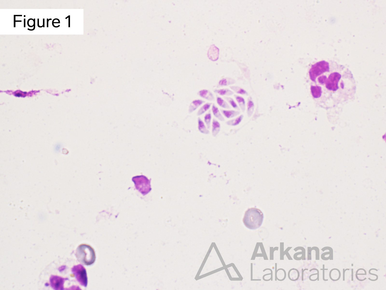
Small collection of T. gondii organism, likely representing a bradyzoites disrupted during the process of performing the intraoperative cytology preparation (“squash prep”)
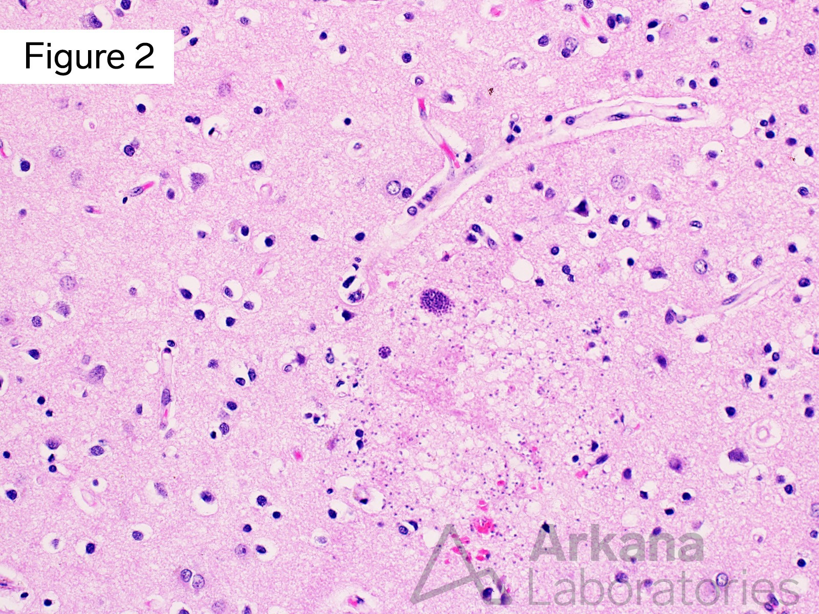
Lower power magnification showing a small area of parenchymal necrosis with two bradyzoites and many free tachyzoites.
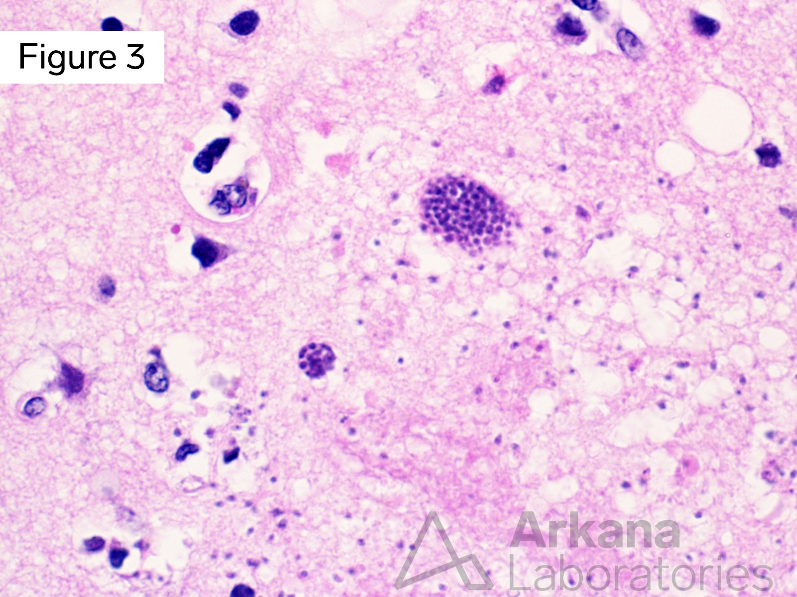
Higher power magnification showing a small area of parenchymal necrosis with two bradyzoites and many free tachyzoites.
Answer: Toxoplasmosis
The intraoperative squash prep and tissue sections show bradyzoites (encysted organisms) and tachyzoites ( individual free organisms) of Toxoplasma gondii. These can be morphologically recognized on H&E. Immunohistochemical staining for the organism is very useful when there is a paucity of organisms or if there is extensive necrosis making it difficult to identify the tachyzoites.
T. gondii is a very successful parasite, infecting approximately 1/3 of the world’s human population. Cats (and other felidae) are definitive hosts, while humans and a wide variety of other warm-blooded animals are intermediate hosts.
In immunocompetent humans primary infection is most commonly asymptomatic to mildly symptomatic, and establishes lifelong latent infection. Reactivation of a previously latent infection occurs in the setting of immunocompromise. Primary or reactivated infection during pregnancy can result in severe fetal encephalitis.
Please see reference for additional information.
Reference(s) / additional reading:
Halonen SK, Weiss LM. Toxoplasmosis. Handb Clin Neurol. 2013;114:125-45. doi: 10.1016/B978-0-444-53490-3.00008-X. PMID: 23829904; PMCID: PMC4157368.
Schlüter D, Barragan A. Advances and Challenges in Understanding Cerebral Toxoplasmosis. Front Immunol. 2019 Feb 14;10:242. doi: 10.3389/fimmu.2019.00242. PMID: 30873157; PMCID: PMC6401564.
Dunay IR, Gajurel K, Dhakal R, Liesenfeld O, Montoya JG. Treatment of Toxoplasmosis: Historical Perspective, Animal Models, and Current Clinical Practice. Clin Microbiol Rev. 2018 Sep 12;31(4):e00057-17. doi: 10.1128/CMR.00057-17. PMID: 30209035; PMCID: PMC6148195.
Attias M, Teixeira DE, Benchimol M, Vommaro RC, Crepaldi PH, De Souza W. The life-cycle of Toxoplasma gondii reviewed using animations. Parasit Vectors. 2020 Nov 23;13(1):588. doi: 10.1186/s13071-020-04445-z. PMID: 33228743; PMCID: PMC7686686.
Quick note: This post is to be used for informational purposes only and does not constitute medical or health advice. Each person should consult their own doctor with respect to matters referenced. Arkana Laboratories assumes no liability for actions taken in reliance upon the information contained herein.