Clinical History:
Patient presented with progressive proximal weakness since August and severe x2 weeks.
Past medical history: hyperthyroidism, hyperlipidemia, herpes simplex, hypertension.
Medications: statin (discontinued 5 days before presentation), tirzepatide, bupropion, levothyroxine, and acyclovir.
Lab results: highest CK 15,991, elevated AST/ALT, WBC 14.7, CRP 0.5, ESR 14, general serologies negative (ANA negative, Jo 1 negative, SCL-70 negative, SM/RNP negative, RF negative). Clinical suspicion for myositis versus rhabdomyolysis.
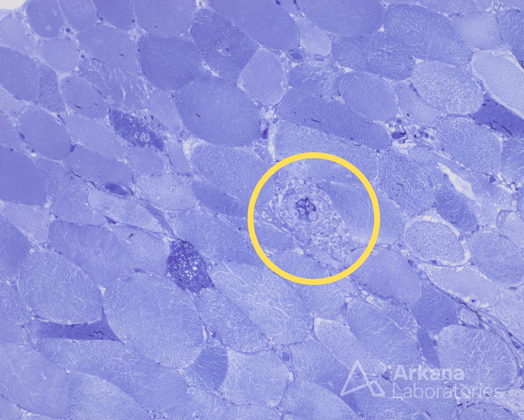
What is in the yellow circle in Figure 1 below?
A. Necrotic fiber
B. Macrophages
C. Lymphocytes
D. Emperipolesis
Here is Figure 1 for reference:
Diagnosis:
Lymphocytes
Lymphocytes surrounding a small vessels as seen on toluidine blue stained 1μm-thick sections.
Quick note: This post is to be used for informational purposes only and does not constitute medical or health advice. Each person should consult their own doctor with respect to matters referenced. Arkana Laboratories assumes no liability for actions taken in reliance upon the information contained herein.