An elderly female in her mid-70’s presents with insidiously progressive severe proximal muscle weakness over the last two to three months to the point that her symptoms of weakness have interfered with her activities of daily living (ADLs) and are also causing her to have limited ambulation. The patient had been on a statin medication for a number of years without issue, and the dosage had recently been increased. Hospital admission showed markedly elevated CPK levels that peaked at CPK ~ 7500 and had moderate downward trend with supportive care. Steroid medication was held until after muscle biopsy of the thigh.
Using the images, which of the following enzymatic histochemical stains is going to provide the neuropathologist with the most information regarding the myopathic change of regenerating muscle fibers?
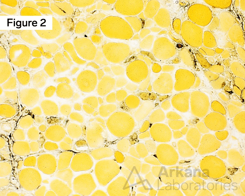
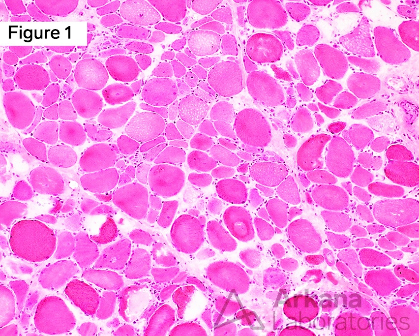
Immune-mediated necrotizing myopathy (IMNM) in an elderly female. Routine frozen hematoxylin and eosin (H&E)-stained sections show frequent slightly basophilic regenerating and pale eosinophilic necrotic muscle fibers with some of the latter undergoing active myophagocytosis (Figure 1). Alkaline phosphatase enzymatic histochemical stain highlights darkly staining variable atrophic regenerating muscle fibers, and also shows minimal endomysial capillary staining, as well as mild patchy perimysial staining (Figure 2). Original magnifications: A. Frozen H&E, 100x; and B. Alkaline phosphatase (also frozen section), 100x.
Frozen H&E Stain
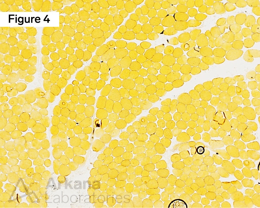
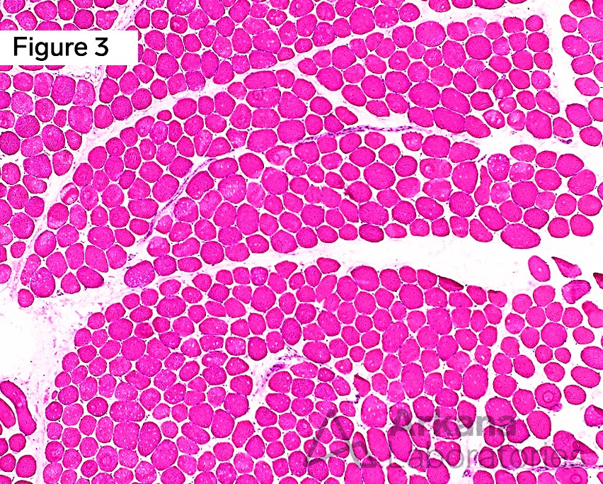
Alkaline phosphatase in normal skeletal muscle for comparison with Figure 2. Routine frozen hematoxylin and eosin (H&E)-stained sections show muscle fibers with minimal variation in muscle fiber size and shape, and are without features of muscle fiber injury (Figure 3). Alkaline phosphatase enzymatic histochemical stain highlights shows minimal endomysial capillary staining, and show no muscle fiber staining or perimysial staining (Figure 4). Original magnifications: A. Frozen H&E, 100x; and B. Alkaline phosphatase (also frozen section), 100x.
Answer: Alkaline Phosphatase
The enzymatic histochemical stain that will help the neuropathologist identify regenerating muscle fibers is alkaline phosphatase. Alkaline phosphatase has many other uses, including aiding in the diagnosis of acquired inflammatory myopathies (i.e. strong perimysial staining in dermatomyositis).
Alkaline phosphatase is an enzyme primarily found in cell membranes where active transport processes occur, such as the endothelium of arterioles and endomysial capillaries. Activity is at the level of the organelles, such as the endoplasmic reticulum, Golgi apparatus, as well as in pinocytotic vesicles.
The alkaline phosphatase reaction is negative in non-injured muscle fibers, but staining is seen in some regenerating and/or non-innervated myofibers. Staining may also be seen in focal necrotic muscle fibers.
Introduction to Alkaline Phosphatase use in human striated skeletal muscle:
Alkaline phosphatase is a generic name for phosphomonoesterases that hydrolyze orthophosphate at an alkaline pH. These enzymes are widely distributed, usually activated by magnesium, manganese, zinc, and cobalt ions, and inhibited by cysteine, cyanides, and arsenates. This is a simultaneous coupling azo dye method first developed in 1944. It has been modified repeatedly. Sodium α-naphthyl acid phosphate, the substrate used in this protocol, is hydrolyzed and then coupled to the diazonium salt (Fast Blue RR) which is then precipitated at the site of enzyme activity.
Source: https://neuromuscular.wustl.edu/pathol/histol/AlP.pdf
Why were the other answers wrong?
Modified Gomori Trichrome stain is one of the most useful special stains employed in all of neuromuscular pathology, as it can aid in identifying mitochondrial content (i.e. ragged red fibers), autophagic conditions (i.e. rimmed vacuoles), and abnormal inclusions (i.e. nemaline rods).
NADH-TR preparation is useful to evaluate internal myofiber architecture, such as target/targetoid muscle fibers, cores, and ragged blue fibers.
The combined SDH-COX preparation is a very useful stain that can provide much information about the muscle fiber’s mitochondrial content, mitochondrial dysfunction/dysregulation, and oxidative phosphorylation stress level. Selective COX-negative/deficient muscle fibers may be seen in a wide variety of conditions, including primary mitochondrial myopathies, increased numbers in sporadic Inclusion Body Myositis (SIBM), and a few scattered negative fibers may also be seen in the context of normal aging (i.e. commensurate with age).
References
Dubowitz V, Sewry CA, Oldfors A. Muscle Biopsy: A Practical Approach, 4th ed. Saunders Elsevier, London, United Kingdom; 2013.
Dubowitz V, Sewry CA, Oldfors A. Muscle Biopsy: A Practical Approach, 5th ed. Saunders Elsevier, London, United Kingdom; 2021.
Cai C, Anthony DC, Pytel P. A pattern-based approach to the interpretation of skeletal muscle biopsies. Mod Pathol. 2019 Apr;32(4):462-483. PMID: 30401945.
Nix JS, Moore SA. What Every Neuropathologist Needs to Know: The Muscle Biopsy. J Neuropathol Exp Neurol. 2020 Jul 1;79(7):719-733. PMID: 32529201.
Alkaline phosphatase. Washington Univeristy Neuromuscular website. https://neuromuscular.wustl.edu/pathol/histol/AlP.pdf
Quick note: This post is to be used for informational purposes only and does not constitute medical or health advice. Each person should consult their own doctor with respect to matters referenced. Arkana Laboratories assumes no liability for actions taken in reliance upon the information contained herein.