
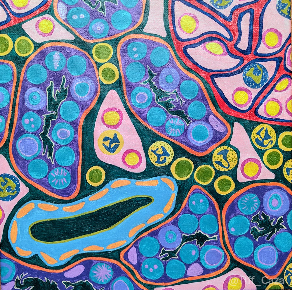
The painting above shows acute tubular injury, reactive changes in tubular epithelial cells, tubulitis, inclusions within tubular epithelium, and interstitial inflammation. These are morphologic changes that can be seen in polyomavirus nephritis. BK virus is the most common etiology of polyomavirus nephritis, while JC virus and simian virus 40 (SV40) are less common etiologies. All are DNA viruses that are non-enveloped and show tropism for the genitourinary tract, especially urothelium (Lusco et al, 2016). Polyomavirus nephritis is a serious complication affecting approximately 5 to 6 percent of kidney transplants, often as a result of over-immunosuppression. The prevalence is higher in patients with ABO-incompatible transplants (18%) and in recipients who are highly sensitized and who underwent desensitization in the peri-transplant period (20%, Nickeleit et al, 2018). Polyomavirus nephritis has serious implications, associated with a high rate of graft loss in transplant recipients.
Polyomavirus nephritis can present clinically with slowly deteriorating renal function with a rise in serum creatinine, cystitis, hemorrhagic cystitis, or rarely ureteral obstruction (Lusco et al, 2016). It is often caused by reactivation of latent virus within infected urothelial or tubular epithelial cells. Latency of BK virus in the genitourinary tract is present in 70 to 90% of individuals and reactivation can occur in an immunosuppressed host.
Risk factors for development of polyomavirus nephritis include older age (>50 years), male gender, Caucasian ethnicity, BK virus negative status prior to transplant, CMV co-infection, diabetes mellitus, and use of triple immunosuppression (prednisone, tacrolimus, mycophenolate mofetil). Patients with HLA mismatches, prior episodes of acute rejection (and methylprednisolone pulse treatment), and calcineurin inhibitor toxicity are at increased risk. Viral factors can also play a role. These include viral serotype, mutations in the non-coding control region, viral capsid protein 1 mutations, and host polymorphisms that affect T lymphocyte production of interferon-gamma in response to viral infection.
A definitive diagnosis of polyomavirus nephritis requires a renal biopsy. Screening for BK viremia is recommended monthly for the first 9 months post-transplant, followed by every 3 months for at least 2 years following transplant if all serum viral PCR studies are negative (Hirsch et al, 2019). BK viremia and BK viruria can be used to monitor disease activity, with >10,000 copies/mL in serum being indicative of activity. Asymptomatic shedding of BK virus within the genitourinary tract can occur, so BK viruria does not necessarily indicate active BK infection and is seen in 30-50% of kidney transplant recipients, yet could decrease with viral clearance (Nickeleit et al, 2018). On a kidney biopsy, SV40 immunohistochemistry increases the sensitivity and specificity for detection of polyomavirus. Nuclear expression of SV40 can be identified in cells without viral cytopathic effects, such as nuclear enlargement or inclusions, so it is recommended for all allograft biopsies.
Polyomavirus nephritis is separated into 3 classes by the Banff classification of polyomavirus nephropathy (Nickeleit et al, 2018). In polyomavirus nephritis class I, less than 1% of tubular epithelial cells show viral replication (by SV40 immunohistochemistry). For polyomavirus nephritis class II, >1% of cells but less than 10% of tubular epithelial cells show SV40 positivity with less than 50% of tubulointerstitial scarring, or >10% of tubular epithelial cells are SV40 positive with less than 10% tubulointerstitial scarring. In polyomavirus class III, there is >10% of tubular epithelial cells with active viral replication with >10% interstitial fibrosis. Classification has prognostic implications, with a 16% rate of graft loss in class I, 31% in class II, and >50% in class III (Nickeleit et al, 2018). Detection of T cell mediated rejection is impaired in the setting of polyomavirus nephritis, as similar histopathologic changes occur with cellular rejection and polyomavirus infection (i.e. tubulitis and interstitial inflammation). One exception is presence of vascular rejection. Since intimal arteritis is not associated with polyomavirus infection, its presence would indicate concurrent rejection.
Treatment of polyomavirus nephritis is generally aimed at immune restoration, with reduction of immunosuppressive therapy. However, this increases the risk of allograft rejection. Intravenous immunoglobulin therapy, leflunomide, cidofovir, or switching immunosuppressive agents can also be attempted in BK nephritis treatment, but there are no randomized controlled clinical trials to date to assess their efficacy (Hirsch et al, 2019).
References:
Hirsch HH, Randhawa PS, AST Infectious Diseases Community of Practice. BK Polyomavirus in Renal Transplant Recipients – Guidelines from the American Society of Transplantation Infectious Diseases Community of Practice. Clin Transplant 2019 Mar 12; e13528.
Lusco MA, Fogo AB, Najafian B, Alpers CE. AJKD Atlas of Renal Pathology: Polyomavirus Nephropathy. Am J Kidney Dis 2016; 68 )6): e37-e38.
Nickeleit V, Singh HK, Randhawa P, Drachenberg CB, Bhatnagar R, Bracamonte E, Chang A, Chon WJ, Dadhania D, Davis VG, Hopfer H, Mihatsch MJ, Papadimitriou JC, Schaub S, Stokes MB, Tungekar MF, Seshan SV. The Banff Working Group Classification of Definitive Polyomavirus Nephropathy: Morphologic Definitions and Clinical Correlations. J Am Soc Nephrol 2018 Feb; 29 (2): 680-693.
Quick note: This post is to be used for informational purposes only and does not constitute medical or health advice. Each person should consult their own doctor with respect to matters referenced. Arkana Laboratories assumes no liability for actions taken in reliance upon the information contained herein.
