Clinical History
- This 40-year old patient with lumbar back pain. MRI demonstrated an intradural extramedullary mass at the level of L3-L4, clinically suspicious for shwannoma. Intraoperatively, the lesion was well-circumscribed and a gross total resection was accomplished. Microscopically, a rare mitotic figure was seen; the proliferative fraction was low; and only very sparse peripheral GFAP reactivity was seen.
The morphologic and immunophenotypic features demonstrated in figures #1 – #4 are consistent with which of the following?
A. Paraganglioma
B. Ependymoma
C. CENET
D. Schwannoma
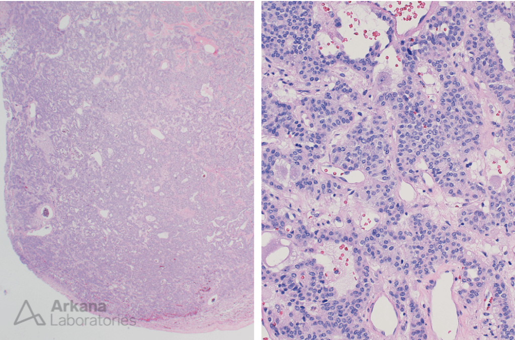

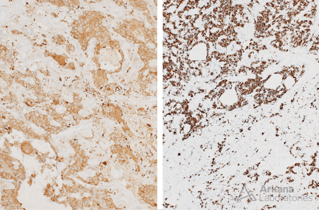

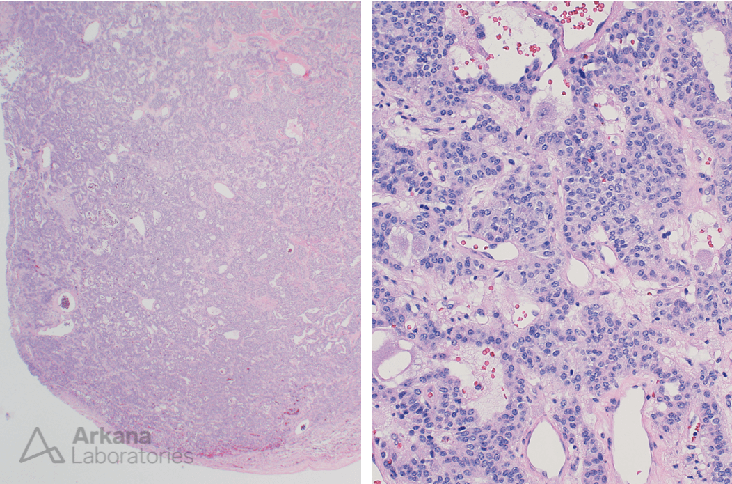
Figure 1: Low and high power H&E
Low magnification image showing a well circumscribed mass.
Medium magnification image showing a cellular proliferation of monotonous appearing epithelioid cells arranged in cords and nests. Scattered relatively mature appearing ganglion cells are present (arrows)
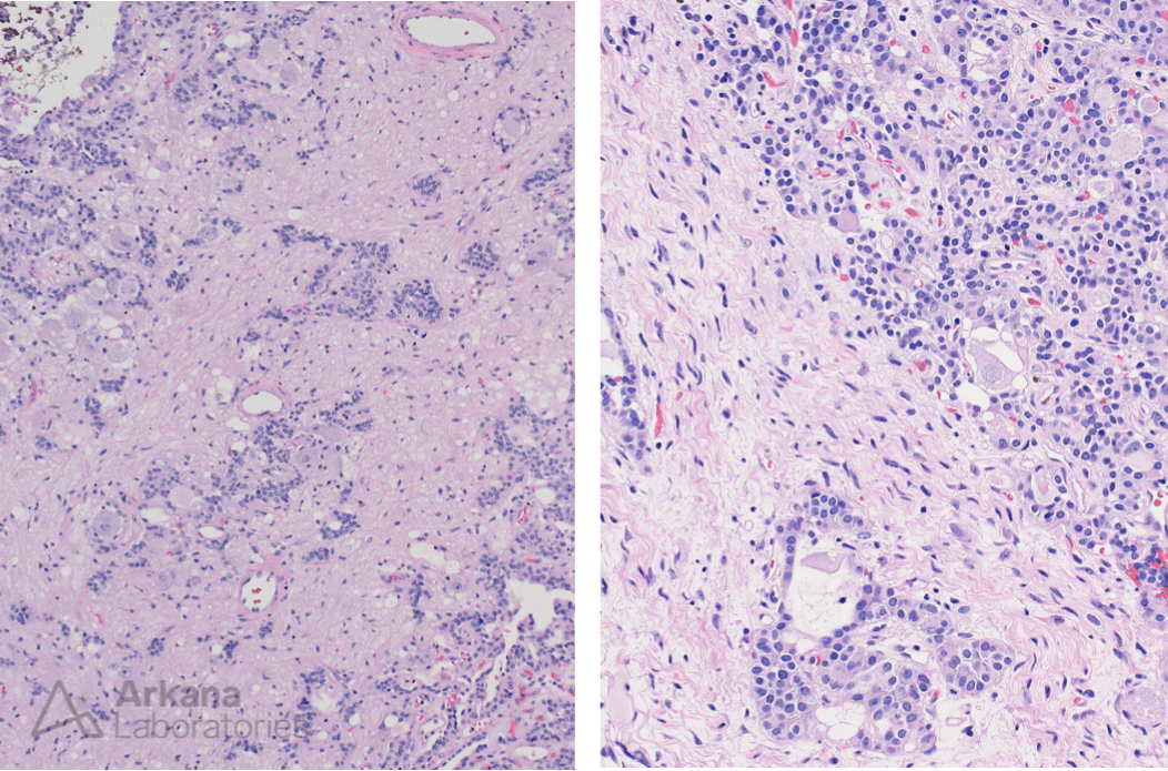
Figure 2: Low and medium magnification H&E
Low and medium magnification images showing ganglioneuromatous component (ganglion cells [arrows] and areas with schwannian type elements [asterix]).
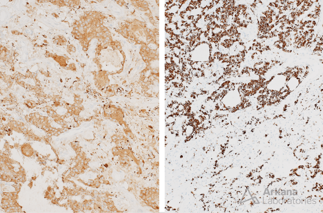
Figure 3: Synaptophysin and low molecular weight cytokeratin IHC
The lesional cells show positive cytoplasmic staining for synaptophysin and low molecular weight cytokeratin.
The lesional cells also showed positive cytoplasmic staining for chromogranin (not shown in the supplied images).
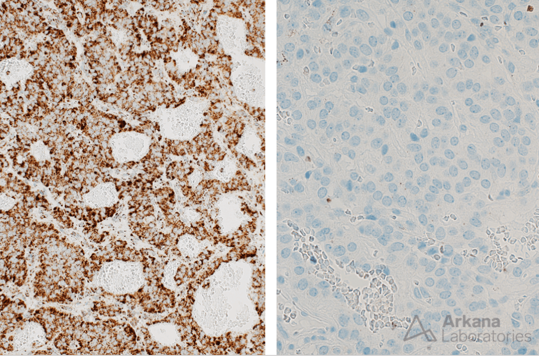
Figure 4: SDHB and GATA3 IHC
The lesional cells showed positive (i.e. retained) staining for SDHB and absence of staining for GATA3.
Correct Answer: C. CENET
- The correct answer is CENET (cauda equina neuroendocrine tumor).
- These are rare neoplasms recently elucidated to be distinct from paragangliomas. They are considered to represent WHO grade 1 lesions (please see references).
Reference(s) / additional reading:
- Asa SL, Mete O, Schüller U, Ramani B, Mirchia K, Perry A. Cauda Equina Neuroendocrine Tumors: Distinct Epithelial Neuroendocrine Neoplasms of Spinal Origin. Am J Surg Pathol. 2023 Apr 1;47(4):469-475. doi: 10.1097/PAS.0000000000002009. Epub 2022 Dec 22. PMID: 36543154.
- Schweizer L, Thierfelder F, Thomas C, Soschinski P, Suwala A, Stichel D, Wefers AK, Wessels L, Misch M, Kim HY, Jödicke R, Teichmann D, Kaul D, Kahn J, Bockmayr M, Hasselblatt M, Younsi A, Unterberg A, Knie B, Walter J, Al Safatli D, May SA, Jödicke A, Ntoulias G, Moskopp D, Vajkoczy P, Heppner FL, Capper D, Hartmann W, Hartmann C, von Deimling A, Reuss DE, Schöler A, Koch A. Molecular characterization of CNS paragangliomas identifies cauda equina paragangliomas as a distinct tumor entity. Acta Neuropathol. 2020 Dec;140(6):893-906. doi: 10.1007/s00401-020-02218-7. Epub 2020 Sep 14. PMID: 32926213; PMCID: PMC7666289.
Quick note: This post is to be used for informational purposes only and does not constitute medical or health advice. Each person should consult their own doctor with respect to matters referenced. Arkana Laboratories assumes no liability for actions taken in reliance upon the information contained herein.