
A 68-year-old man presents with insidiously progressive proximal left thigh weakness with atrophy, left foot drop, and right hand grip strength weakness. Laboratory work-up reveals elevated CPK 850 and negative myositis panel. A muscle biopsy of the left thigh is performed, and shows a polymyositis-type injury pattern with myofibers containing rare rimmed vacuoles. Using the movie below, what characteristic finding is seen with Congo red stain in this case?
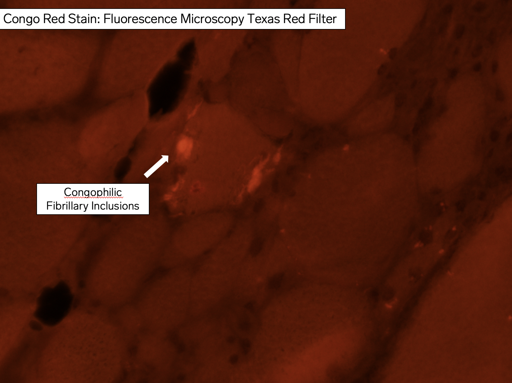
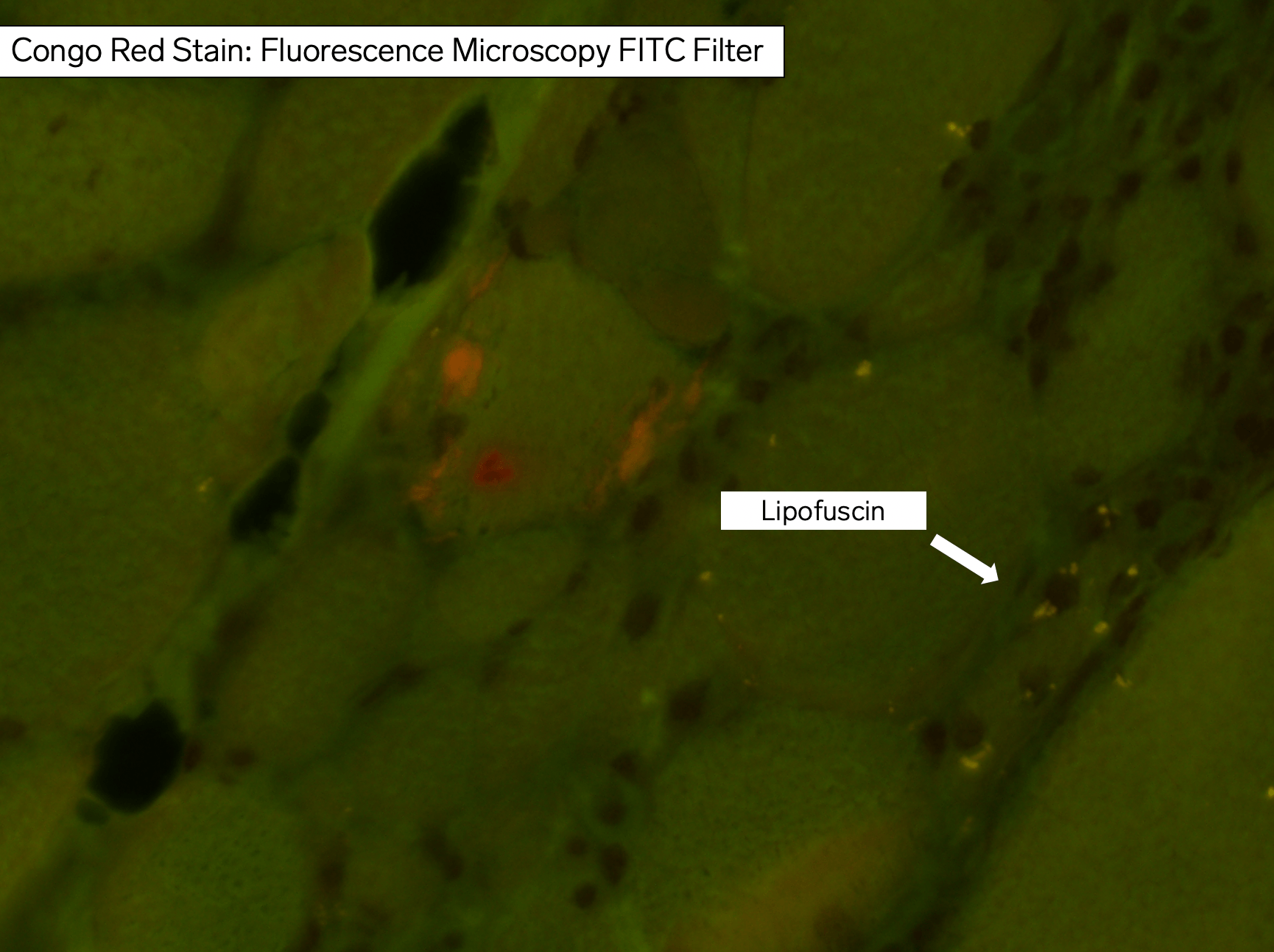
Answer: The images show different modalities examining a Congo red special histochemical stain demonstrating characteristic congophilic inclusions of sporadic Inclusion Body Myositis (sIBM). With a Congo red stain, the skein-like inclusions of sIBM are bright red under UV light using a Texas Red excitation filter (fluorescence microscopy with rhodamine optics), transition to orange under UV light with a FITC filter, and appear pale orange-pink with routine light microscopy. To learn more about sIBM, see the reference below.
Why were the other answers incorrect?
A mild amount of lipofuscin pigment deposition is very common in adult skeletal muscle biopsies. Increased amounts may be seen in elderly patients, and nonspecifically in other myopathies, such as late (adult) onset acid maltase deficiency (Pompe disease, GSD II). As seen in the movie, Congo red stain yields lipofuscin as white-yellow with FITC under UV light, and is granular dark orange under routine light microscopy.
The characteristic finding of Nemaline rod myopathy, nemaline rods, is not observed in this case. The clinical vignette does not fit this congenital myopathy, although an adult variant is described.
Desmin protein aggregates may be seen in myofibrillar myopathy (i.e. desminopathy) and myofibrillar myopathies may show myofibrillar disorganization with ill-defined flocculent sarcoplasmic congophilia. However, this finding is not the one demonstrated in this case.
References:
Greenberg SA. Inclusion body myositis: clinical features and pathogenesis. Nat Rev Rheumatol. 2019 May;15(5):257-272. PMID: 30837708.
Quick note: This post is to be used for informational purposes only and does not constitute medical or health advice. Each person should consult their own doctor with respect to matters referenced. Arkana Laboratories assumes no liability for actions taken in reliance upon the information contained herein.