This 50-year-old patient presented with complaints of slowly progressive proximal lower extremity muscle weakness of multiple year duration. Laboratory studies showed normal CPK.
Which of the following best characterizes the features seen in Figures #1 and #2?
A. Contraction bands
B. Normal muscle
C. Rimmed vacuoles
D. Necrotic fibers

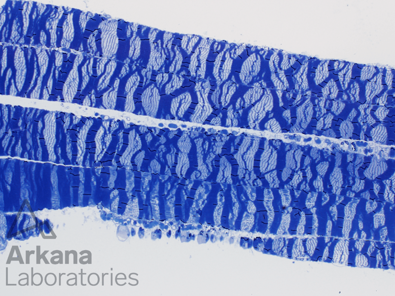
Low magnification image showing alternating dark and pale bands.
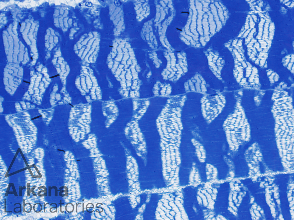
High magnification image showing alternating dark and pale bands. These represent alternating areas of contracted and stretched-out sarcomeres, respectively.
Which of the following best characterizes the features seen in Figures #1 and #2?
A. Contraction bands < Correct Answer
B. Normal muscle
C. Rimmed vacuoles
D. Necrotic fibers
The irregular thick “striped” appearance represents contraction bands. This artifact is commonly seen in muscle biopsies samples exposed to excessive saline during transportation from the operating room to the pathology lab.
Quick note: This post is to be used for informational purposes only and does not constitute medical or health advice. Each person should consult their own doctor with respect to matters referenced. Arkana Laboratories assumes no liability for actions taken in reliance upon the information contained herein.