What would be your leading diagnosis in a patient with elevated ACE levels and hilar lymphadenopathy?
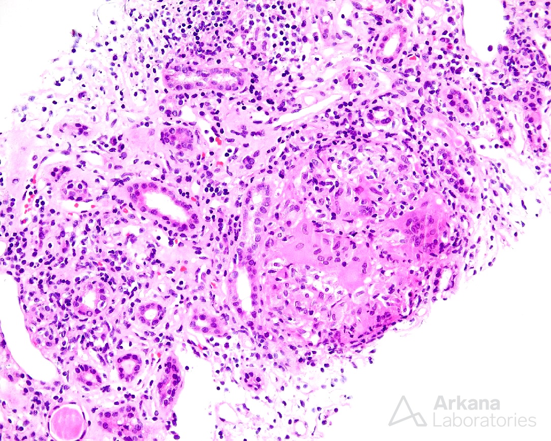
The light microscopic image depicts a non-necrotizing granuloma which, in the clinical context, would be compatible with Sarcoidosis. Histologic features of Sarcoidosis typically include well-formed non-caseating granulomas composed of epithelioid histiocytes and multinucleated giant cells along with an active tubulointerstitial nephritis typically composed of a lymphoplasmacytic infiltrate, rarely containing eosinophils. Interestingly, rare cases of this disease have shown a concomitant glomerular disease, the most common being Membranous nephropathy. Of important note, the biopsy findings of a non-necrotizing granulomatous tubulointerstitial nephritis are not specific to Sarcoidosis and the differential diagnosis would also include drug/hypersensitivity reaction (most common), vasculitis, and infection (i.e. mycobacterial, fungal).
Quick note: This post is to be used for informational purposes only and does not constitute medical or health advice. Each person should consult their own doctor with respect to matters referenced. Arkana Laboratories assumes no liability for actions taken in reliance upon the information contained herein.