Clinical History:
This 65-year-old patient presented with recent onset of seizures. Brain MRI demonstrated a single large ring enhancing mass within the left frontal lobe. The mass was resected.
Question:
Cytomorphologic features seen in Figures #1 and #2 are most consistent with which of the following?
A. Astrocytic neoplasm
B. Metastatic neoplasm
C. Normal brain
D. Infection
Answer:
Cytomorphologic features seen in Figures #1 and #2 are most consistent with which of the following?
A. Astrocytic neoplasm <– correct answer
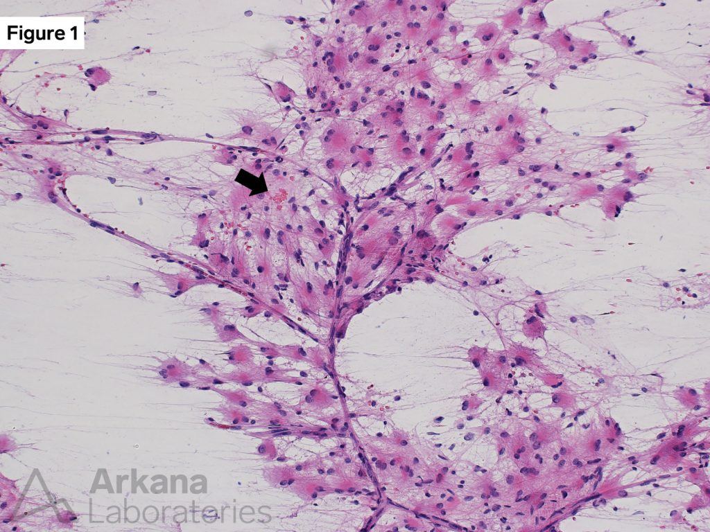
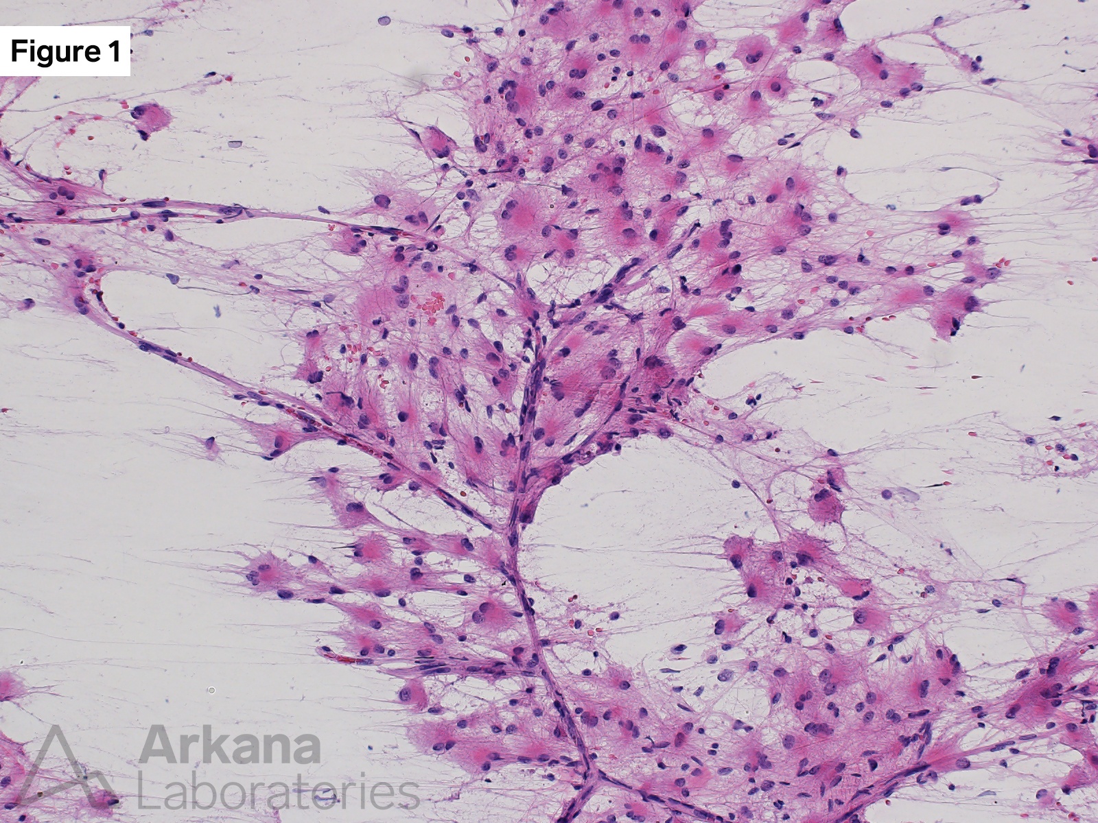
Figure 1: H&E stained cytology preparation 200x original magnification
Medium magnification image of the intraoperative “squash prep” showing atypical glial cells with enlarged nuclei (compare to the occasional RBCs indicated by arrow in this image), small to medium sized bellies of brightly eosinophilic cytoplasm, and fine cytoplasmic processes. The cytomorphologic features are astrocytic in appearance.
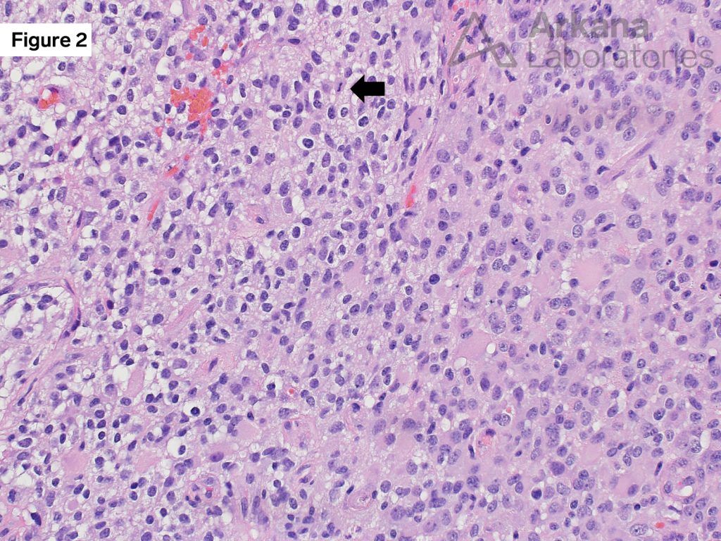
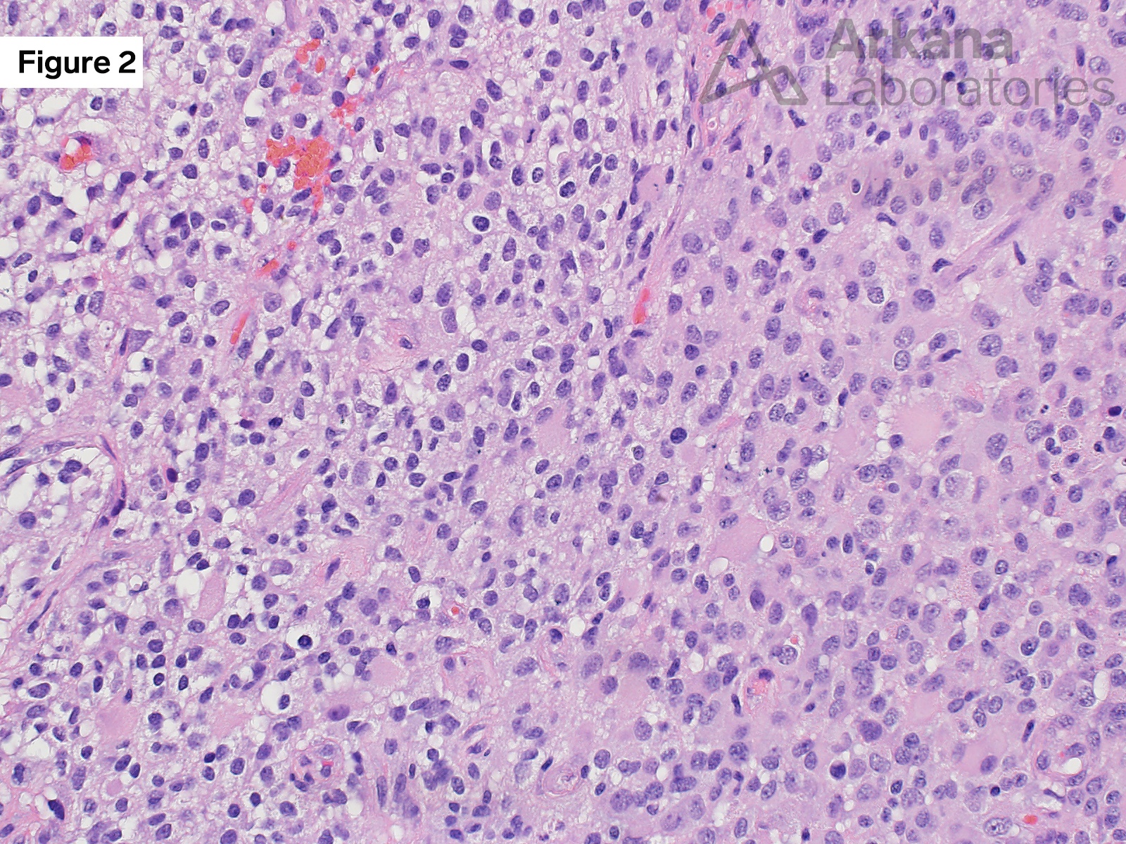
Figure 2: H&E stained FFPE section 200x original magnification
Medium magnification image of the permanent FFPE section showing the atypical astrocytic appearing cells. A single mitotic figure is seen in this image (see arrow).
This represents an astrocytic neoplasm.
Quick note: This post is to be used for informational purposes only and does not constitute medical or health advice. Each person should consult their own doctor with respect to matters referenced. Arkana Laboratories assumes no liability for actions taken in reliance upon the information contained herein.