What does modified gomori trichrome (MGT) show?
Considering the clinical vignette from the prior week (elderly male with proximal muscle weakness). Staining the skeletal muscle biopsy in combination with Modified Gomori Trichrome (MGT) and oxidative stains can provide the neuropathologist with much information, including clues as to the ultrastructural abnormality of this myopathy.
Using the images below, True or False: The Modified Gomori Trichrome (MGT) staining in this skeletal muscle biopsy is definitive for mitochondria.
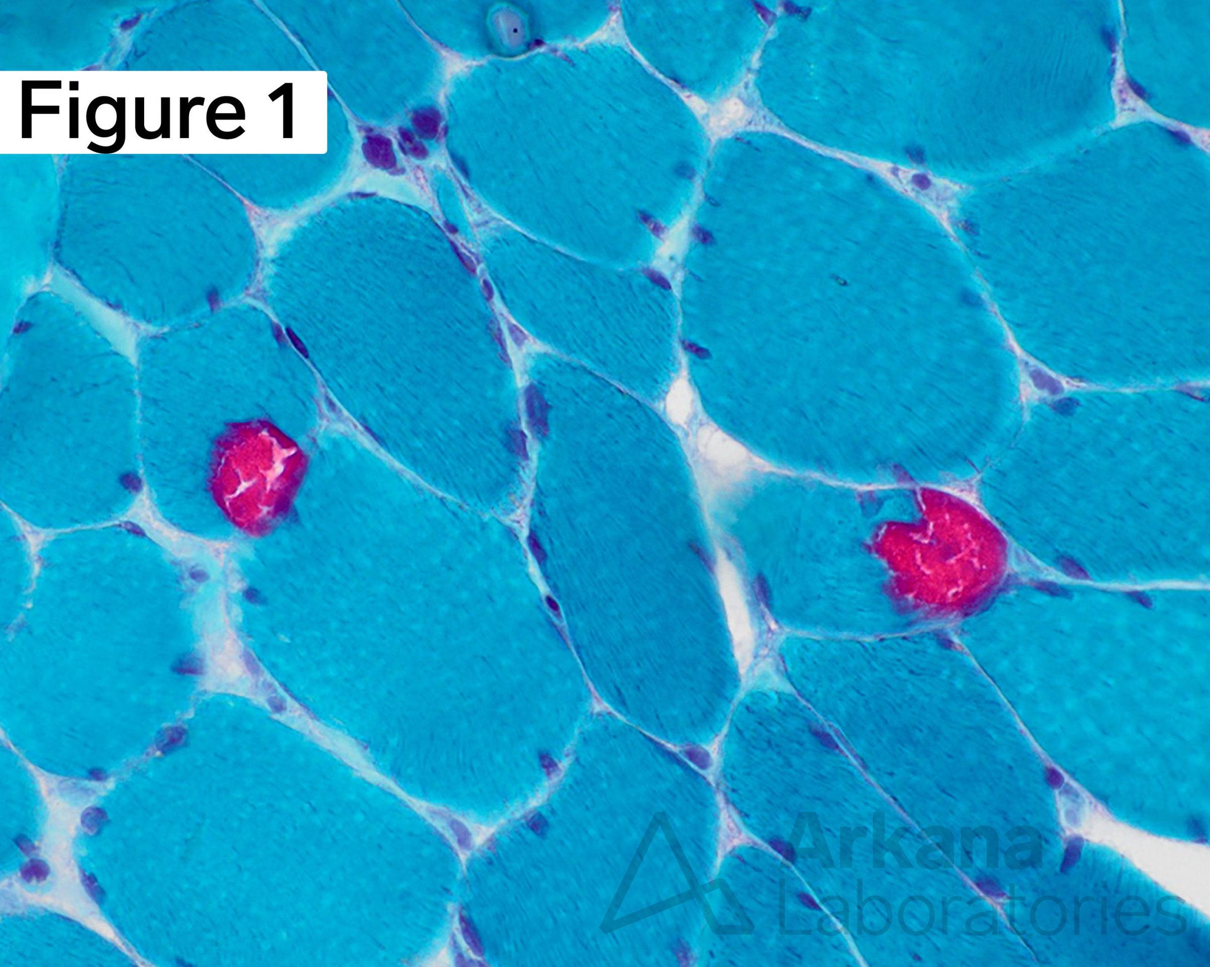
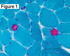
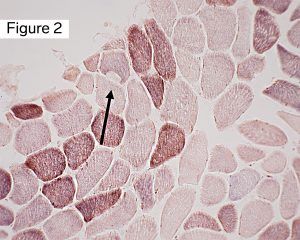
Frozen sections of skeletal muscle with Tubular Aggregate Myopathy (TAM) demonstrating two near by muscle fibers with round to ovoid eccentric and subsarcolemmal inclusion-type material with dense bright fuchsinophilic (red) staining character (1) with Modified Gomori Trichrome (MGT) preparation. A deeper section in the same case shows a similarly shaped subsarcolemmal area devoid of staining (black arrow) with the combined succinate dehydrogenase – cytochrome C oxidase (SDH-COX) preparation (2). (MGT and SDH-COX stains: original magnification 400x and 200x, respectively).
Answer: False
Not all dense bright fuchsinophilic (red) staining with Modified Gomori Trichrome (MGT) preparation yields the ragged red fibers of mitochondrial myopathy, but such staining can be meaningful to the neuropathologist in other ways to deduce the etiology of the myopathic process. In this case, the MGT staining is that of myopathy with tubular aggregates.
The function of oxidative staining with the combined succinate dehydrogenase – cytochrome C oxidase (SDH-COX) preparation is most commonly used for the assessment of mitochondria content (mitochondrial accumulations) and mitochondrial dysfunction (selective COX-negative/deficient blue fibers with SDH-COX). In this case of myopathy with tubular aggregates, the area devoid of staining (black arrow) indicates a paucity of mitochondria in the inclusion material, further shedding insight into the etiology of the MGT staining. We’ll expand further into the nuance of SDH-COX staining in future #NeuroNotes.
Key Point Take-Away:
Not all red staining with Modified Gomori Trichrome (MGT) is mitochondria. When in doubt, stain it out. Combination use of oxidative stains, such as SDH-COX, can further define inclusion material. Ultrastructural examination with electron microscopy may be helpful.
Quick note: This post is to be used for informational purposes only and does not constitute medical or health advice. Each person should consult their own doctor with respect to matters referenced. Arkana Laboratories assumes no liability for actions taken in reliance upon the information contained herein.