Clinical history
This 60-year-old patient presented with proximalmuscle weakness and pain of one-month’s duration. Laboratory studies showed elevated CPK (~12000).
Question:
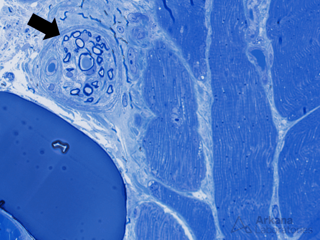
Which of the following does the structure indicated by the arrow in the figure represent?
A. Nerve fascicule
B. Muscle fiber
C. Muscle spindle
D. Blood vessel

Figure 1: Toluidine blue stained thick section 600x original magnification
Answer: A. Nerve fascicle
This high magnification image shows multiple large and smaller diameter myelinated axons (the myelin sheaths are dark blue; while the axons surrounded by the myelin sheaths are very pale blue/white). Note the well-defined sheath formed by perineurial cells surrounding the bundle of axons.
Note the muscle fibers in predominantly longitudinal orientation in the right half of the image, and the adipocyte in the lower left region of the image.
Quick note: This post is to be used for informational purposes only and does not constitute medical or health advice. Each person should consult their own doctor with respect to matters referenced. Arkana Laboratories assumes no liability for actions taken in reliance upon the information contained herein.