Clinical History
- This 60-year-old patient presented with progressive symmetrical subacute onset proximal muscle weakness involving their lower and upper extremities. Electrodiagnostic studies showed myopathic features. Laboratory studies showed marked elevation of CPK. Muscle biopsy was performed to evaluate for myopathy. No myotoxic medications were noted in the patient’s home medication list. They were treated with steroid prior to muscle biopsy and showed some clinical improvement.
Based on Figures #1 – #4 the appearance of the NADH-TR enzyme histochemical stain (Figure #2) is best explained by redistribution of which of the following?
A. Lysosomes
B. Mitochondria
C. Sarcomeres
D. Triads
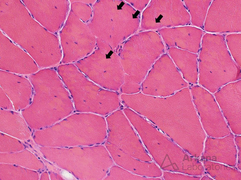
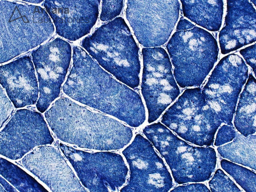
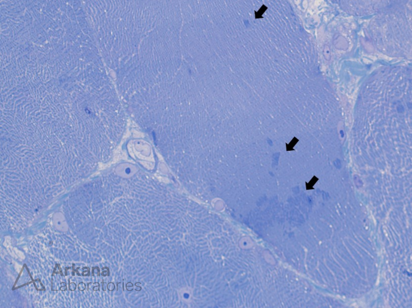
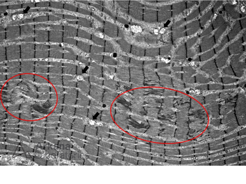
Figure 1: Higher magnification image of H&E stained section showing an increase in the number of muscle fibers with internalized nuclei. In other areas of this patient’s biopsy frequent regenerating and necrotic myofibers were present. Note also the subtle areas of somewhat brighter smudgy areas within the sarcoplasm of multiple myofibers (for examples see arrows).
Figure 2: Higher magnification image of NADH-TR (Nicotinamide adenine dinucleotide tetrazolium reductase) stained section showing frequent myofibers with multiple relatively well-circumscribed areas of staining pallor. These are most obvious in the darker staining Type 1 myofibers, but also appear to be present in the paler staining Type 2 fibers.
Figure 3: High magnification image of toluidine blue stained thick section showing multiple area with darker blue tinctoral quality (see arrows).
Figure 4: Ultrastructural study shows multiple small areas with z-band streaming and some associated disorganization of sarcomeric arrangement (see asterix). Note: that these areas of sarcomeric disorganization (circled) show fewer mitochondria (see arrows) than the surrounding more normal appearing areas.
Correct answer: Mitochondria
- The areas of staining pallor are felt to be due to redistribution of mitochondria out of the areas of sarcomeric disorganization.
- NADH-TR, cytochrome oxidase (COX) and succinic acid dehydrogenase (SDH) enzyme histochemical stains (oxidative stains) highlight mitochondria.
- Areas of oxidative staining pallor may be seen in central core disease (structured and unstructured cores), multiminicore disease, and denervation (target and targetoid fibers).
Quick note: This post is to be used for informational purposes only and does not constitute medical or health advice. Each person should consult their own doctor with respect to matters referenced. Arkana Laboratories assumes no liability for actions taken in reliance upon the information contained herein.