A sural nerve biopsy was performed on this 60-year-old patient to evaluate for mononeuropathy.
What structure is demonstrated in Figures #1 – #4?
A. Glomus body
B. Golgi tendon organ
C. End-stage nerve
D. Pacinian corpuscle
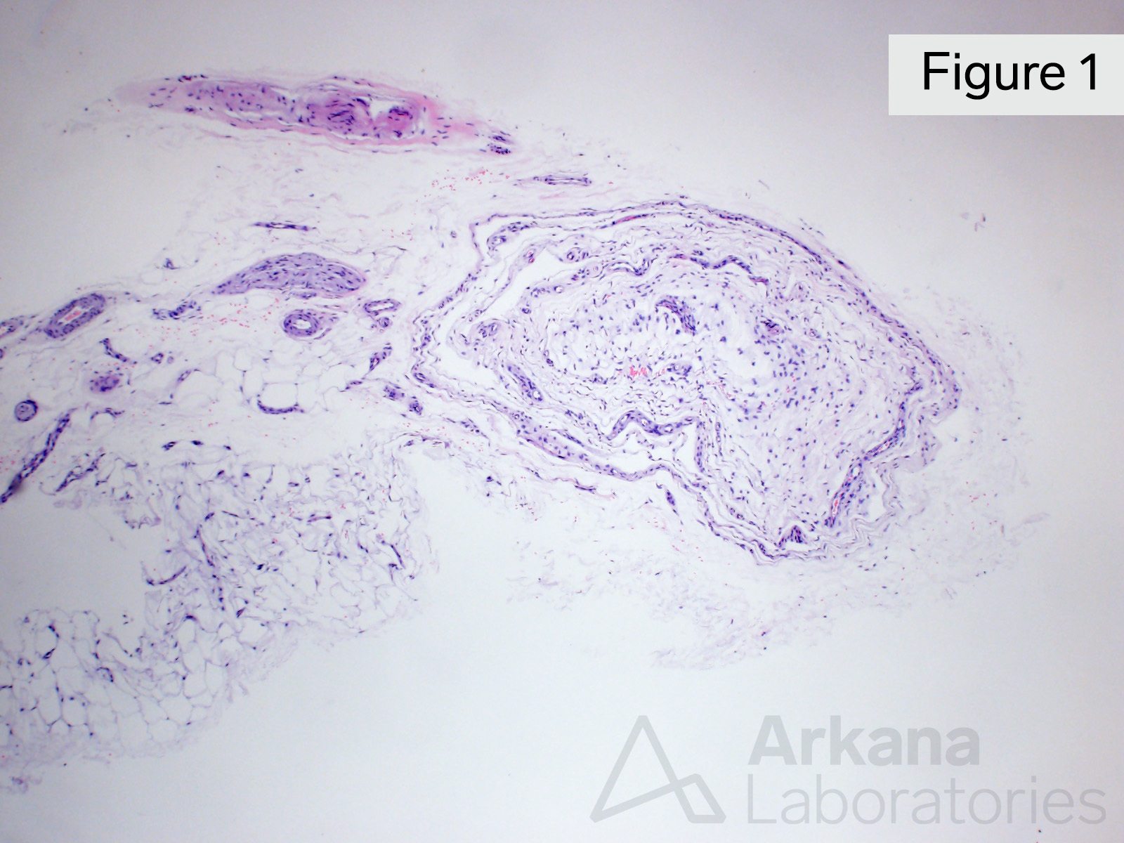
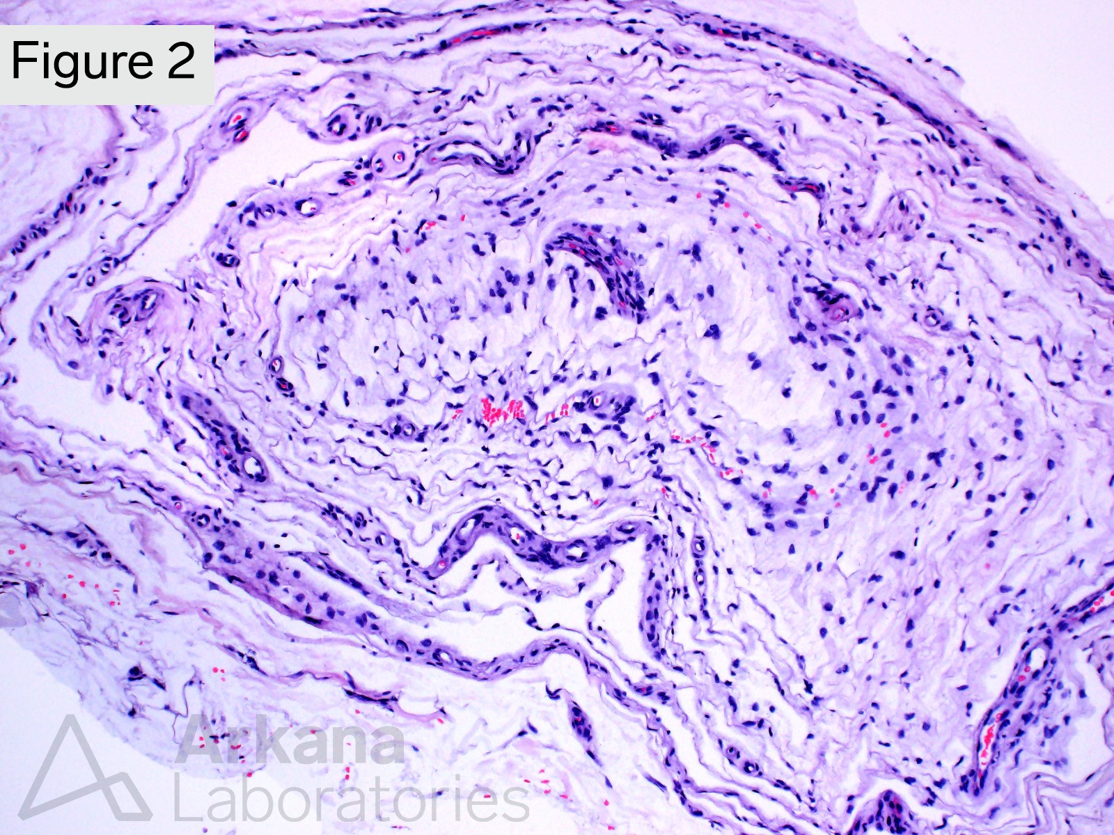
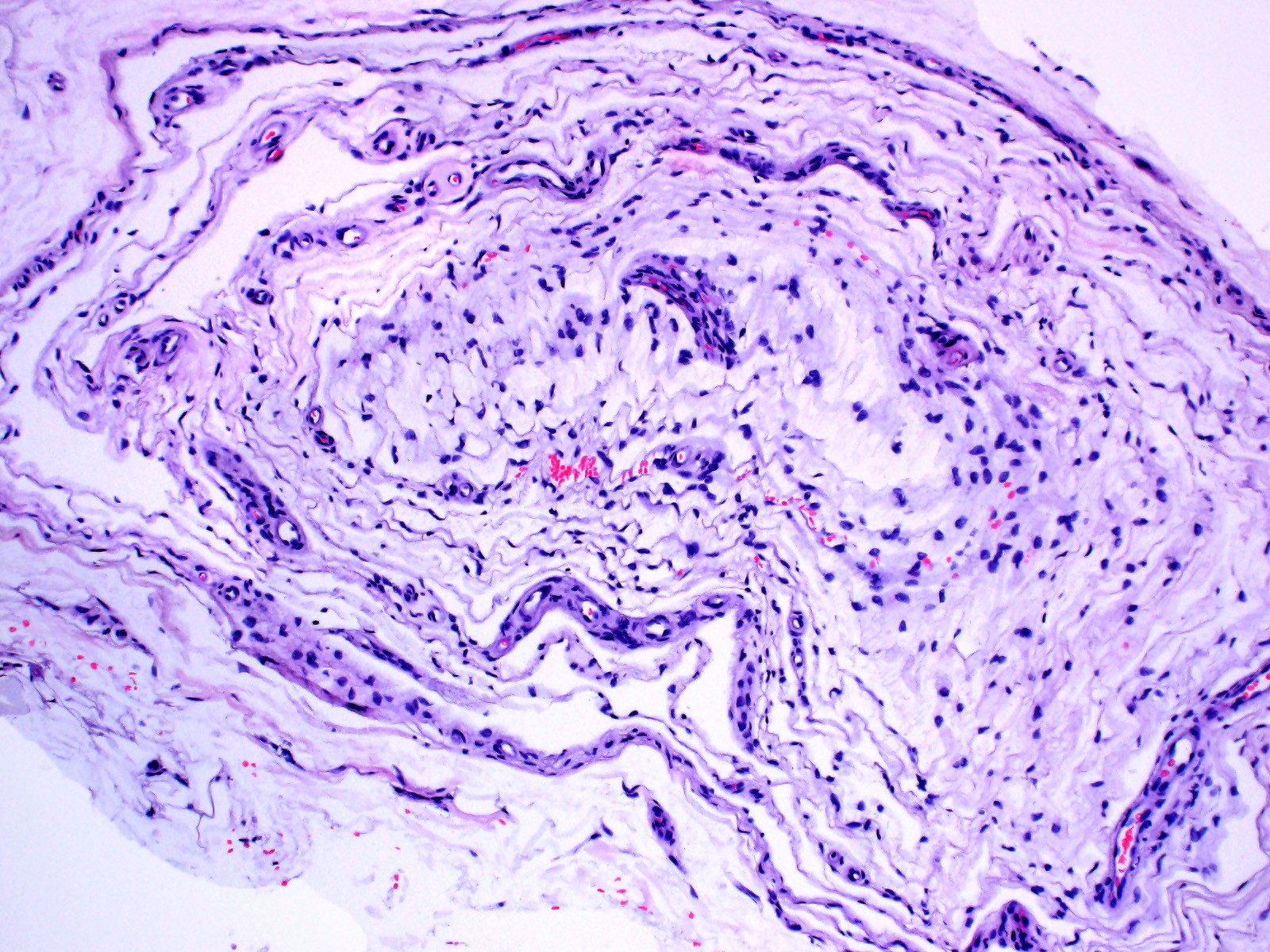
These images show the “onion skin” like layers of a specialized mechanoreceptor. The central nerve ending is not in the plane of section in these images.
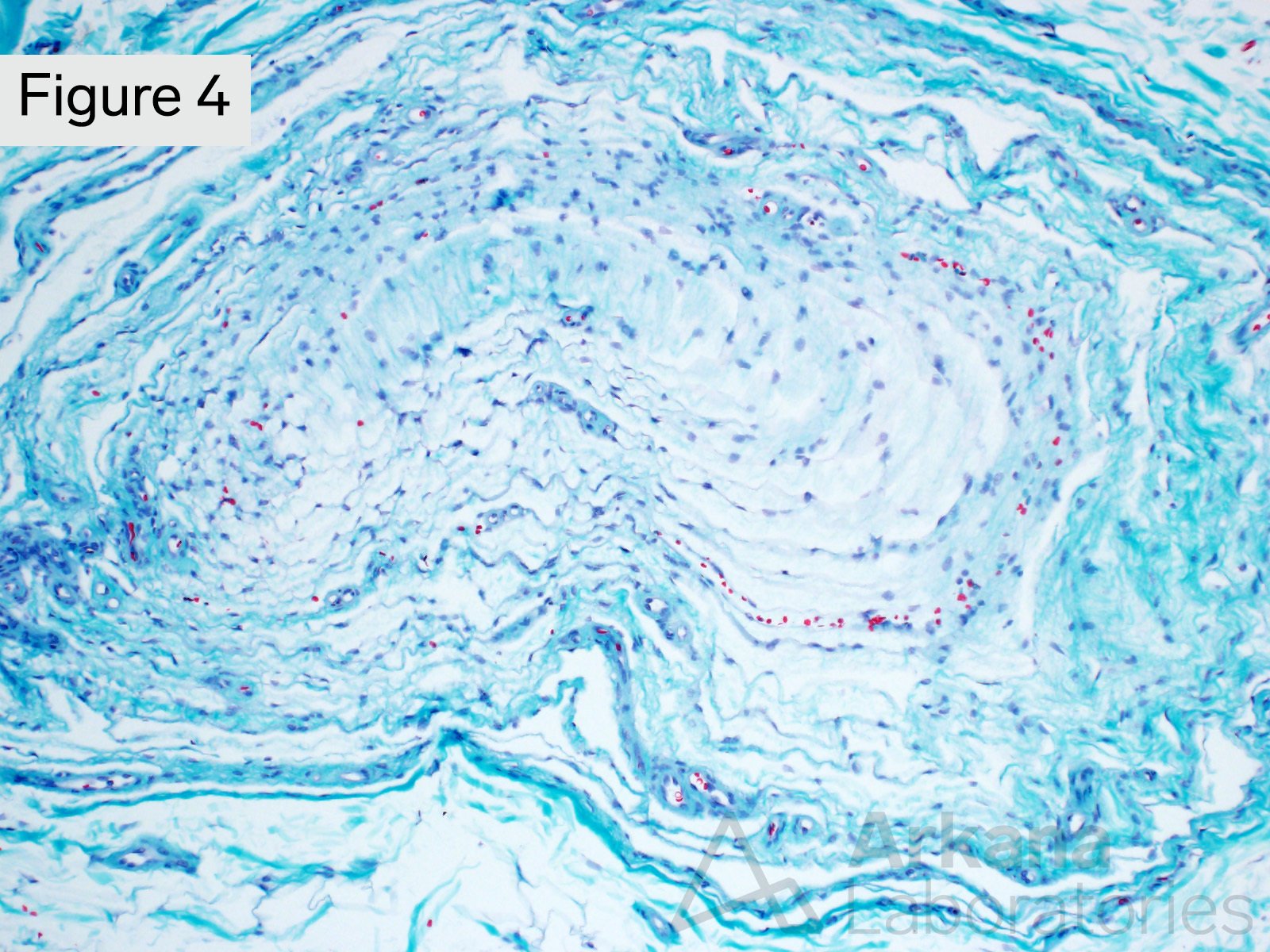
Answer: Pacinian corpuscle
The images show the incidental presence of a Pacinian corpuscle (one type of mechanoreceptor) within the soft tissue adjacent to this patient’s sural nerve. Pacinian corpuscles are involved in vibratory and pressure sensation.
Reference(s) / additional reading:
Suazo I, Vega JA, García-Mesa Y, García-Piqueras J, García-Suárez O, Cobo T. The Lamellar Cells of Vertebrate Meissner and Pacinian Corpuscles: Development, Characterization, and Functions. Front Neurosci. 2022 Mar 9;16:790130. doi: 10.3389/fnins.2022.790130. PMID: 35356056; PMCID: PMC8959428.
Bajwa H, Al Khalili Y. Physiology, Vibratory Sense. 2022 May 8. In: StatPearls [Internet]. Treasure Island (FL): StatPearls Publishing; 2022 Jan–. PMID
https://en.wikipedia.org/wiki/Pacinian_corpuscle : 31194428.
Quick note: This post is to be used for informational purposes only and does not constitute medical or health advice. Each person should consult their own doctor with respect to matters referenced. Arkana Laboratories assumes no liability for actions taken in reliance upon the information contained herein.