Clinical History
The 60-year-old patient was admitted for complaints of acute left lower extremity pain and weakness, and was found to have a left popliteal deep vein thrombosis. Laboratory studies showed elevated CPK (30000). No myotoxic medications were noted in the patient’s home medication list.
Which of the following diagnoses is most consistent with Figures #1 – #3 taken of this patient’s muscle biopsy?
A. Polymyositis
B. Dermatomyositis
C. Ischemic myopathy
D. Vasculitis

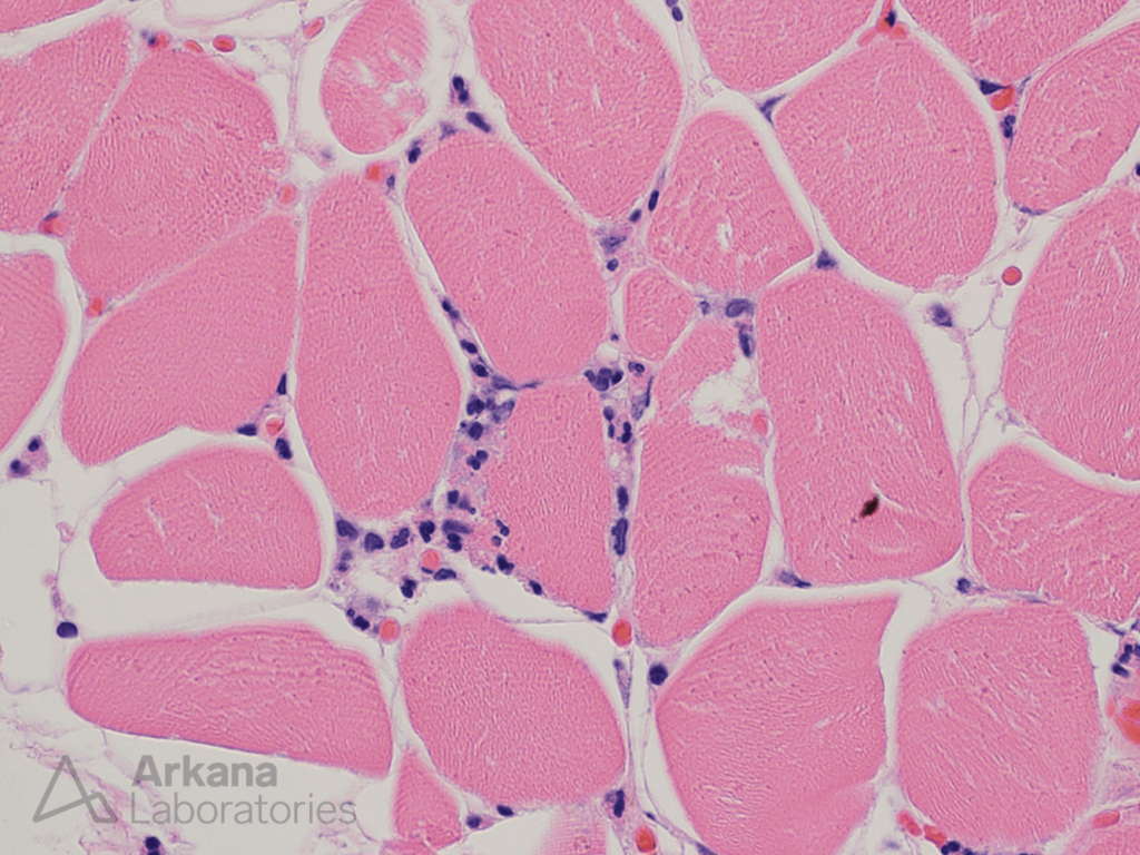
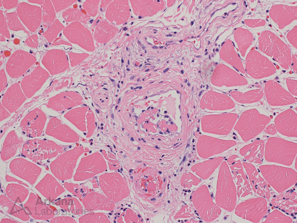
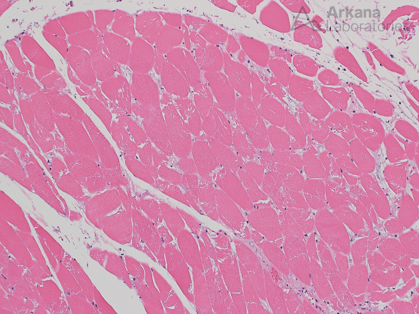
Figure 1: This lower magnification image shows skeletal muscle in cross section. The muscle fibers show diffuse homogeneous mild pallor of staining. Also, note that most of the muscle fibers do not show visibly staining nuclei.
Figure 2: This high magnification image shows focal sparse acute inflammatory cells between the muscle fibers (i.e. within the endomysium). These represent limited acute inflammatory reaction to diffuse muscle fiber injury in the setting of poor vascular perfusion.
Figure 3: This medium magnification image shows the focal presence of intravascular thrombi which have been surfaced by endothelial cells (i.e. early organization of thrombus).
Correct answer: C. Ischemic myopathy
- Ischemic myonecrosis is the best answer.
- Compartment syndrome with associated ischemic myonecrosis has uncommonly been reported in association with deep vein thrombosis.
- Ischemic myonecrosis may also be seen in the setting of peripheral vascular disease in diabetic patients (diabetic myonecrosis).
Quick note: This post is to be used for informational purposes only and does not constitute medical or health advice. Each person should consult their own doctor with respect to matters referenced. Arkana Laboratories assumes no liability for actions taken in reliance upon the information contained herein.