
This 50-year-old patient underwent muscle biopsy to evaluate for cramps and myalgias.
What is the structure in the following figure?
A. Peripheral nerve
B. Muscle spindle
C. Blood vessel
D. Parasite
Correct answer: Peripheral Nerve
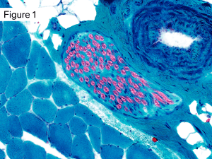
Small fascicle of unremarkable appearing peripheral nerve included within the muscle biopsy.
The structure in the center of the image is a small peripheral nerve fascicle. Modified Gomori Trichrome stain highlights the myelin sheaths. In the cross sectional plane the axon wrapped by myelin can be seen as a central small dark green dot.
Note also: Small muscular artery in the upper right corner. Skeletal muscle fibers in the lower left half of the image
Quick note: This post is to be used for informational purposes only and does not constitute medical or health advice. Each person should consult their own doctor with respect to matters referenced. Arkana Laboratories assumes no liability for actions taken in reliance upon the information contained herein.