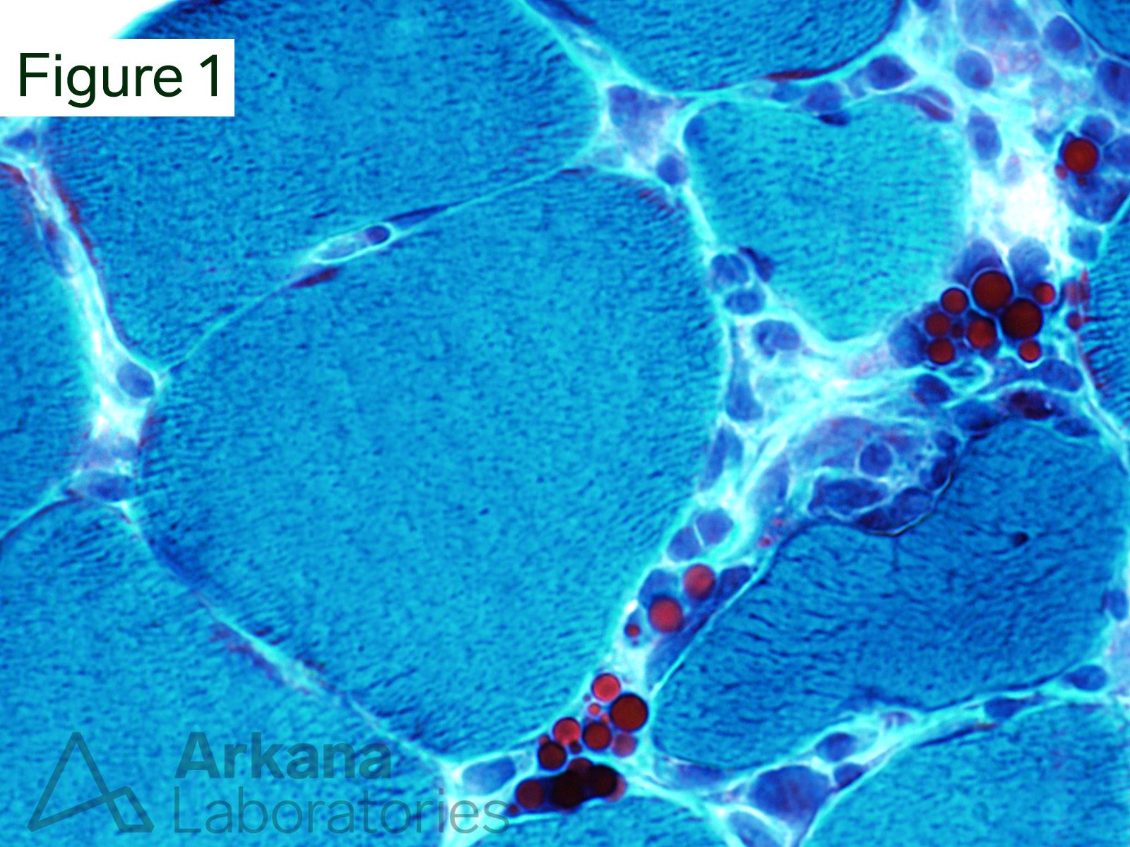
This 50-year-old patient’s muscle biopsy showed morphologic changes of polymyositis. Their past medical history is significant for rheumatoid arthritis.
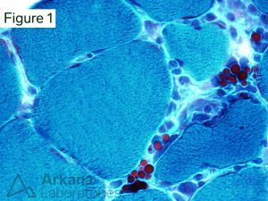
What are the spherical structures seen in Figures #1 and #2?
A. Cryptococcus
B. Psammoma bodies
C. Talc granules
D. Russell bodies
Two peripheral nerve fascicles show diffuse moderate to marked thickening of the perineurium (arrows).
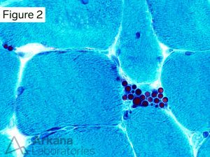
Answer: Russell bodies
The grape-like clusters of spherical inclusions represent accumulation of immunoglobulin (Ig) within the endoplasmic reticulum of plasma cells. These structures are called “Russell bodies”. Plasma cells containing Russell bodies are known as “Mott cells.” Russell bodies and Mott cells may be seen in autoimmune disease, infection, and neoplastic plasma cell proliferations.
Reference(s)/Additional Reading
Iwamoto T, Witmer R. Light and electron microscopy on plasma cells and Russell bodies in the iris of a chronic uveitis patient. Invest Ophthalmol. 1969 Dec;8(6):563-82. PMID: 5359574.
Mossuto MF, Ami D, Anelli T, Fagioli C, Doglia SM, Sitia R. Biochemical nature of Russell Bodies. Sci Rep. 2015 Jul 30;5:12585. doi: 10.1038/srep12585. PMID: 26223695; PMCID: PMC4649990.
Quick note: This post is to be used for informational purposes only and does not constitute medical or health advice. Each person should consult their own doctor with respect to matters referenced. Arkana Laboratories assumes no liability for actions taken in reliance upon the information contained herein.