Clinical History:
This 25-year-old patient with a history of Neurofibromatosis type 1(NF1) was found to have a relatively rapidly enlarging 13 cm posterior chest wall mass. The mass was resected.

Question:
Based on the clinical history and Figures #1 to #4, what is your diagnosis?
Answer:
The correct answer is C. MPNST.
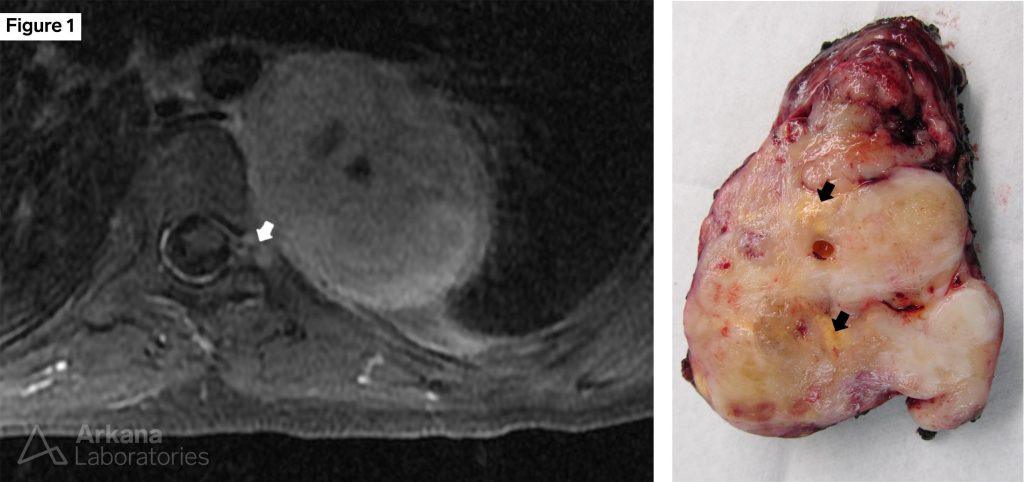
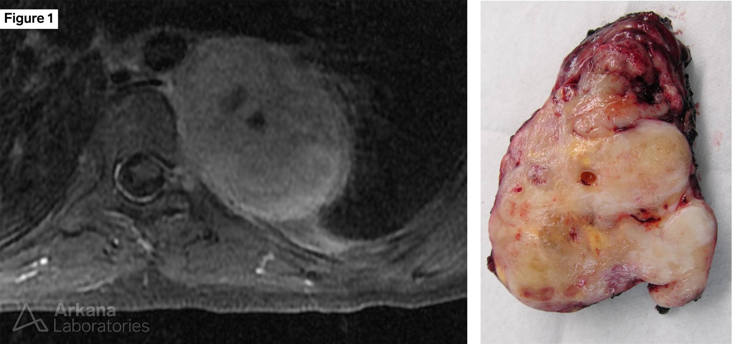
Figure 1: MRI (T1 AXIAL FSE with contrast) and cut surface of resection specimen
Note that the mass appears to extend into the neural foramina (white arrow). The mass showed a multilobulated appearing cut surface with areas of necrosis (black arrows).
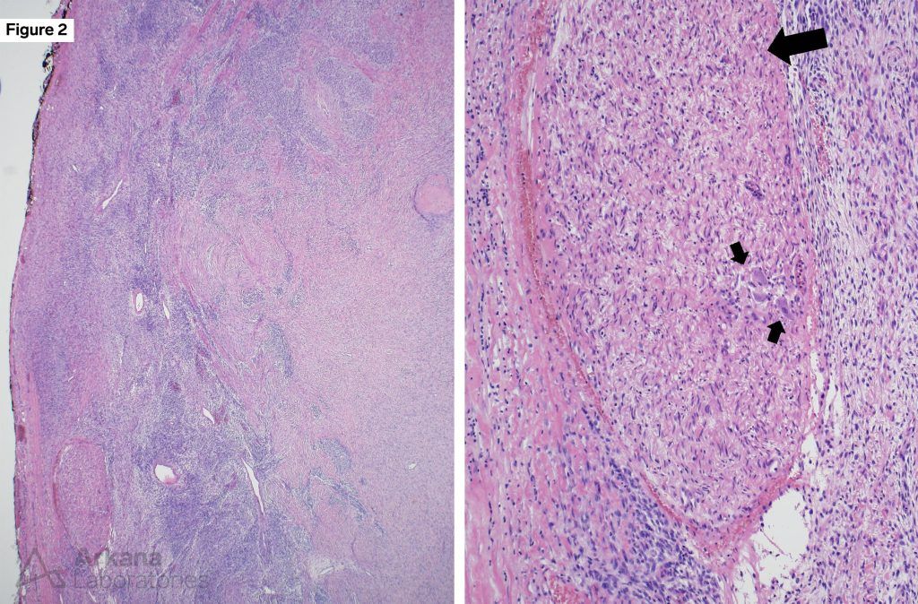
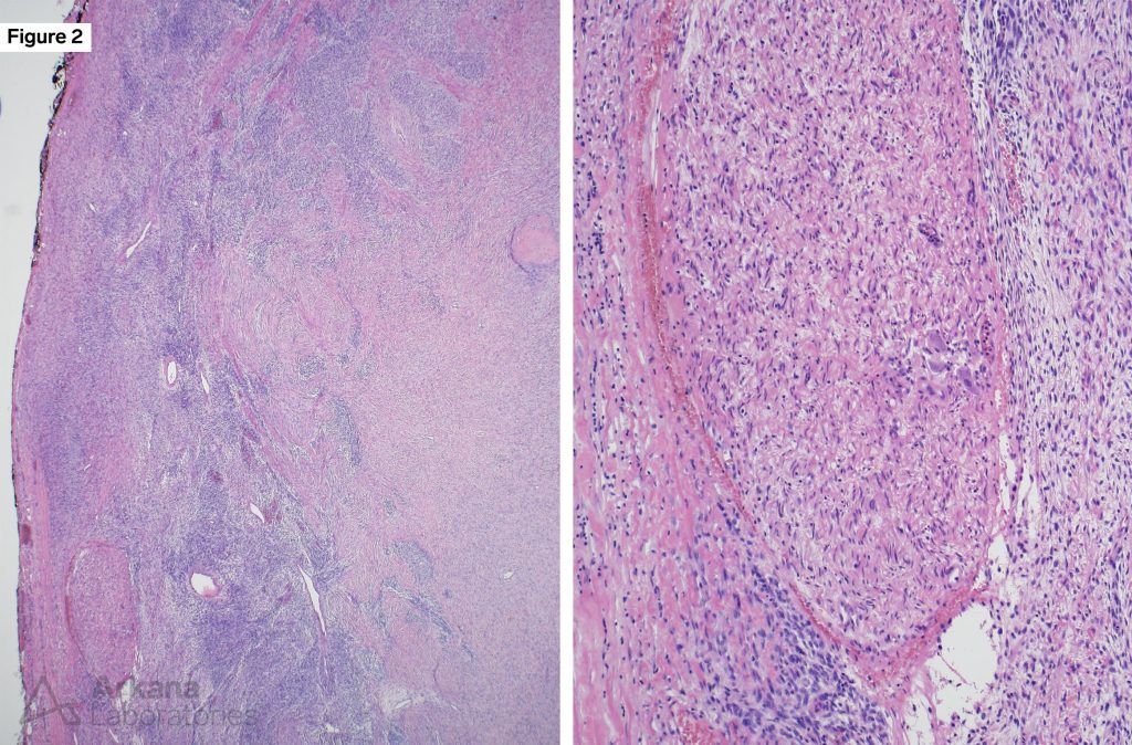
Figure 2: Hematoxylin and eosin stained section
The low power image to the left shows a variably cellular lesion. The higher power image to the right shows an entrapped nerve fascicle (large arrow) surrounded by a variably cellular spindle cell neoplasm. The presence of several mature neurons (small arrows) indicates this likely represents the junction with a ganglion.
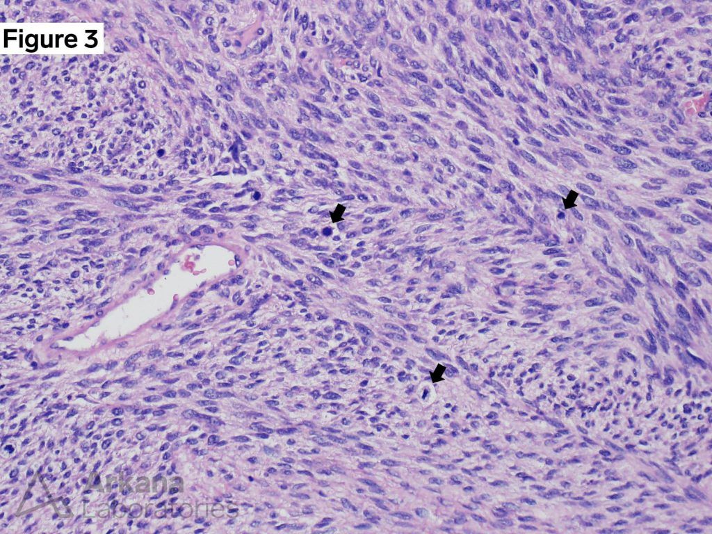
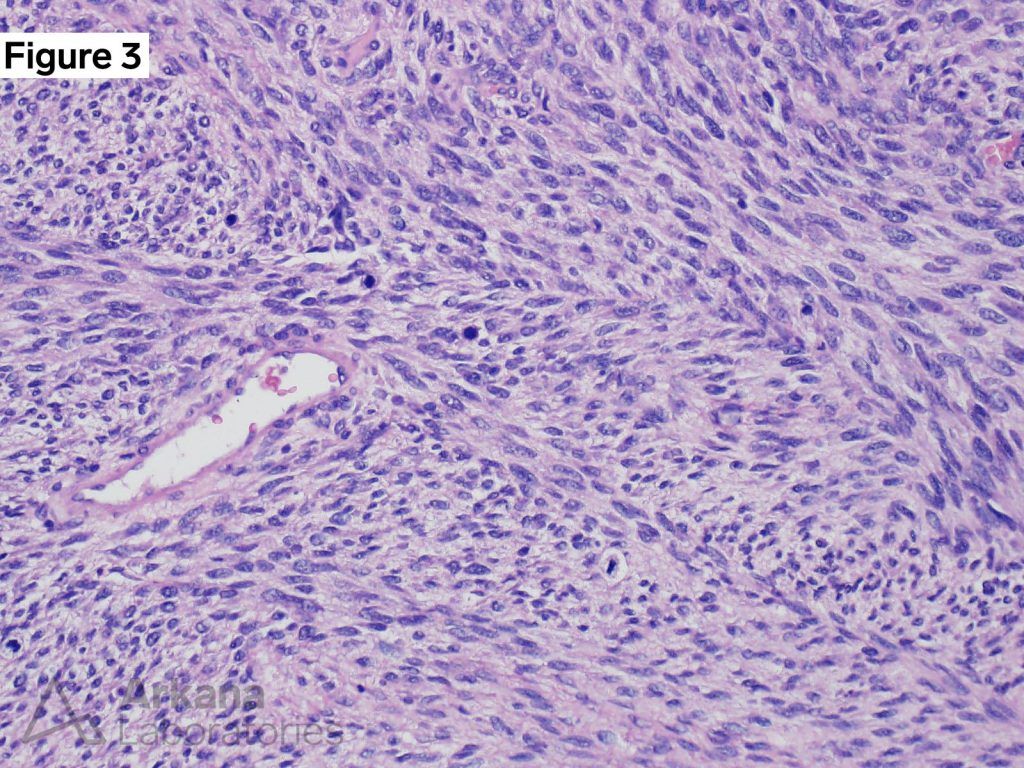
Figure 3: Hematoxylin and eosin stained section
The higher power shows a cellular spindle cell lesion, arranged in a fascicular pattern. Several mitotic figures (arrows) are present in this field, and the lesion had greater than 10 mitoses per 10 hpf.
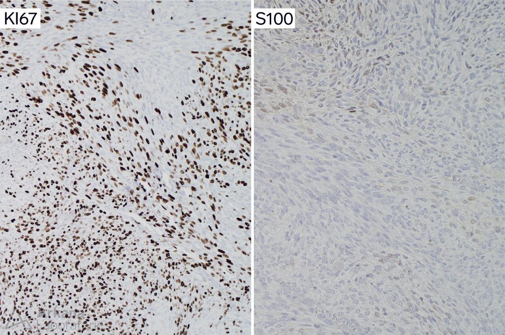
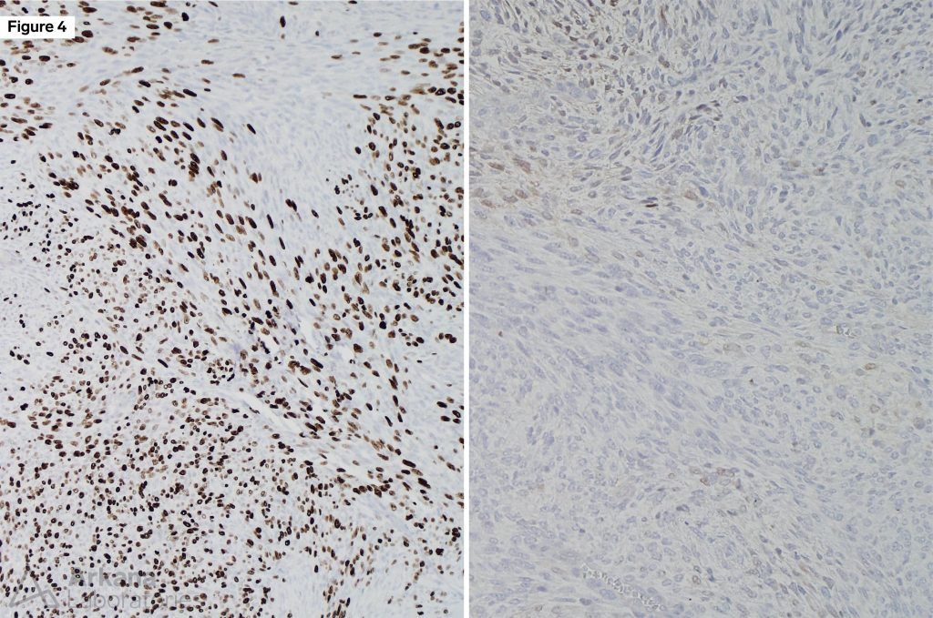
Figure 4: KI67 and S100 protein immunohistochemical stains
The lesional cells showed marked increase in proliferative fraction (left image). There was only patchy weak staining of neoplastic cells for S100 protein (right image).
MPNST
The clinical and morphologic features are consistent with the diagnosis of malignant peripheral nerve sheath tumor (MPNST) arising in a neurofibroma.
Reference(s)/Additional Reading:
Quick note: This post is to be used for informational purposes only and does not constitute medical or health advice. Each person should consult their own doctor with respect to matters referenced. Arkana Laboratories assumes no liability for actions taken in reliance upon the information contained herein.