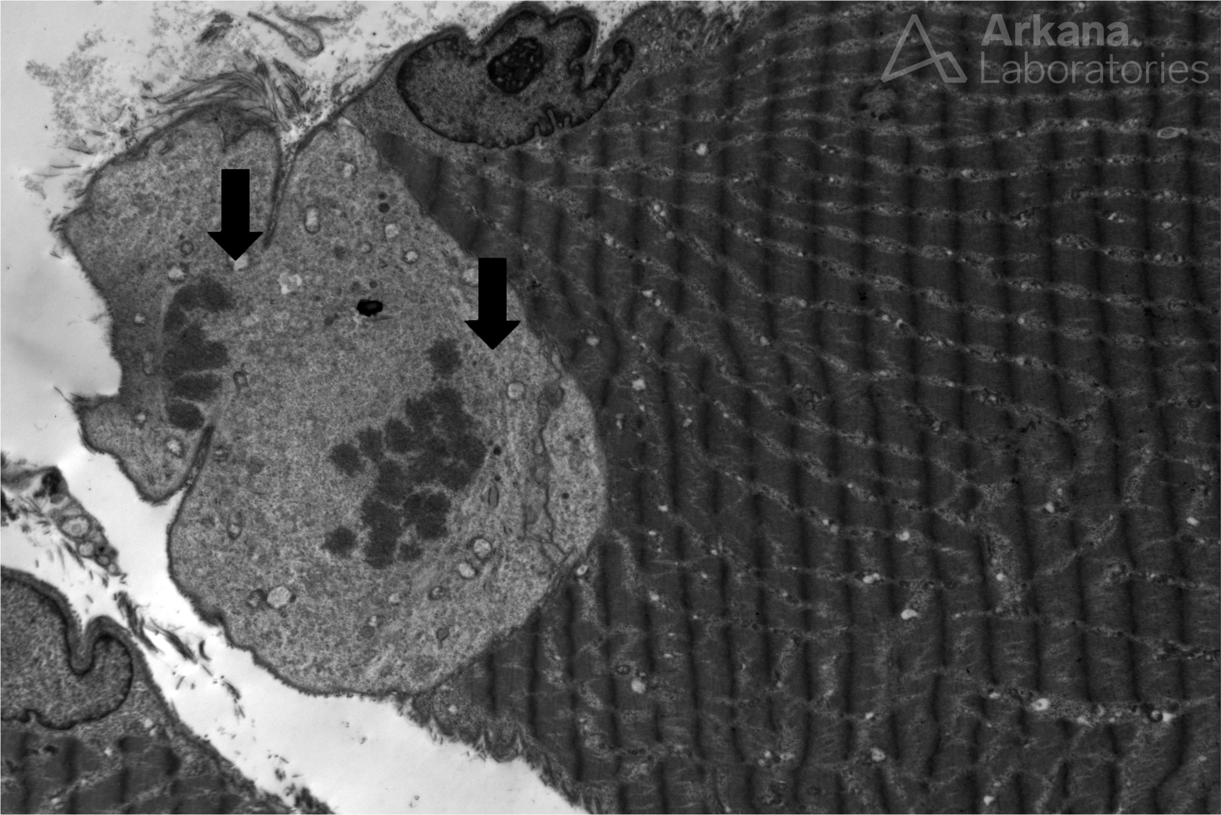

Figure 1: Electron microscopy
Electron microscopy on a muscle biopsy with myopathic changes showed a satellite cell undergoing cellular division. Note the condensed chromosomes (arrows) which have been separated by the mitotic spindle into still conjoined daughter cells.
Muscle fibers represent a post-mitotic syncytium. Satellite cells are separate from myofibers, but are located within the same surrounding basement membrane. Satellite cells fuse with myofibers and are critical in the processes of muscle fiber maintenance, hypertrophy and repair. Please see references.
Reference(s)/Additional Reading:
- Feige P, Brun CE, Ritso M, Rudnicki MA. Orienting Muscle Stem Cells for Regeneration in Homeostasis, Aging, and Disease. Cell Stem Cell. 2018 Nov 1;23(5):653-664. doi: 10.1016/j.stem.2018.10.006. PMID: 30388423; PMCID: PMC6262894.
-
Cossu G, Tajbakhsh S. Oriented cell divisions and muscle satellite cell heterogeneity. Cell. 2007 Jun 1;129(5):859-61. doi: 10.1016/j.cell.2007.05.029. PMID: 17540166.
- Murach KA, Fry CS, Kirby TJ, Jackson JR, Lee JD, White SH, Dupont-Versteegden EE, McCarthy JJ, Peterson CA. Starring or Supporting Role? Satellite Cells and Skeletal Muscle Fiber Size Regulation. Physiology (Bethesda). 2018 Jan 1;33(1):26-38. doi: 10.1152/physiol.00019.2017. PMID: 29212890; PMCID: PMC5866409.
Quick note: This post is to be used for informational purposes only and does not constitute medical or health advice. Each person should consult their own doctor with respect to matters referenced. Arkana Laboratories assumes no liability for actions taken in reliance upon the information contained herein.