This 60-year-old patient presented with complaints of fatigue, lower back pain, and slowly progressive bilateral foot drop. Laboratory studies showed negative/normal anti-MAG antibodies, paraneoplastic autoantibody panel, IFE, and SPEP. EMG demonstrated severe distal denervation in a length-dependent manner, consistent with polyneuropathy. NCS showed features of severe axonal sensorimotor polyneuropathy. On physical examination, the patient was noted to have distal lower extremity muscle weakness and decreased to absent deep tendon reflexes.
What normal structure of the peripheral nerve myelin sheath is indicated by the bracket in Figures #1 and #2?
A. Outer mesaxon
B. Myelin incisure
C. Cajal bands
D. Axoplasm
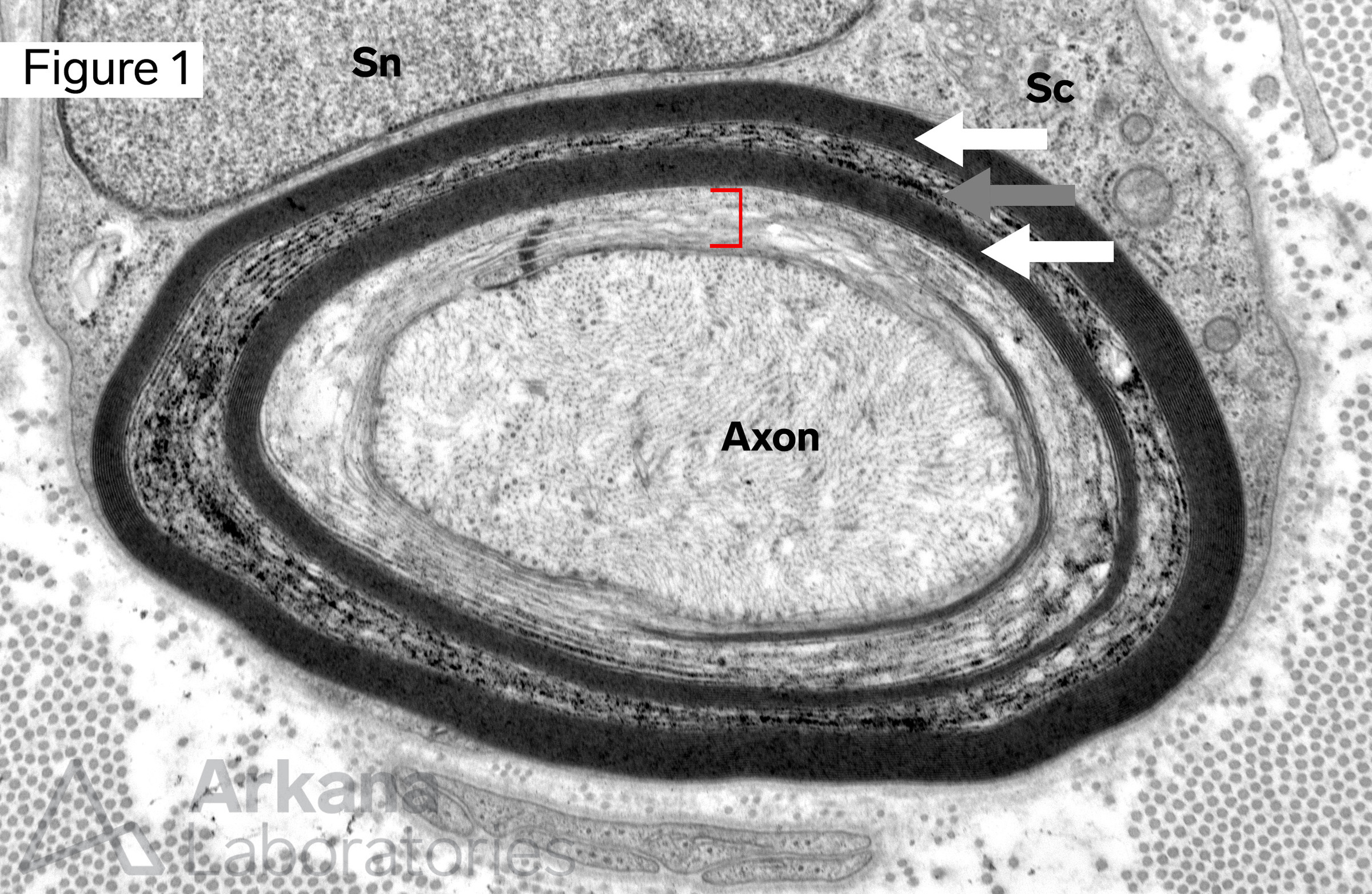
The bracket delineates a circumferential area of the myelin sheath were multiple wraps of the Schwann cell cytoplasm are not compacted. Peripheral to this region there is an area of traction artifact (gray arrow) sandwiched between layers of well-compacted myelin (white arrows). Note the surrounding Schwann cell cytoplasm (Sc) and nucleus (Sn), and axon.
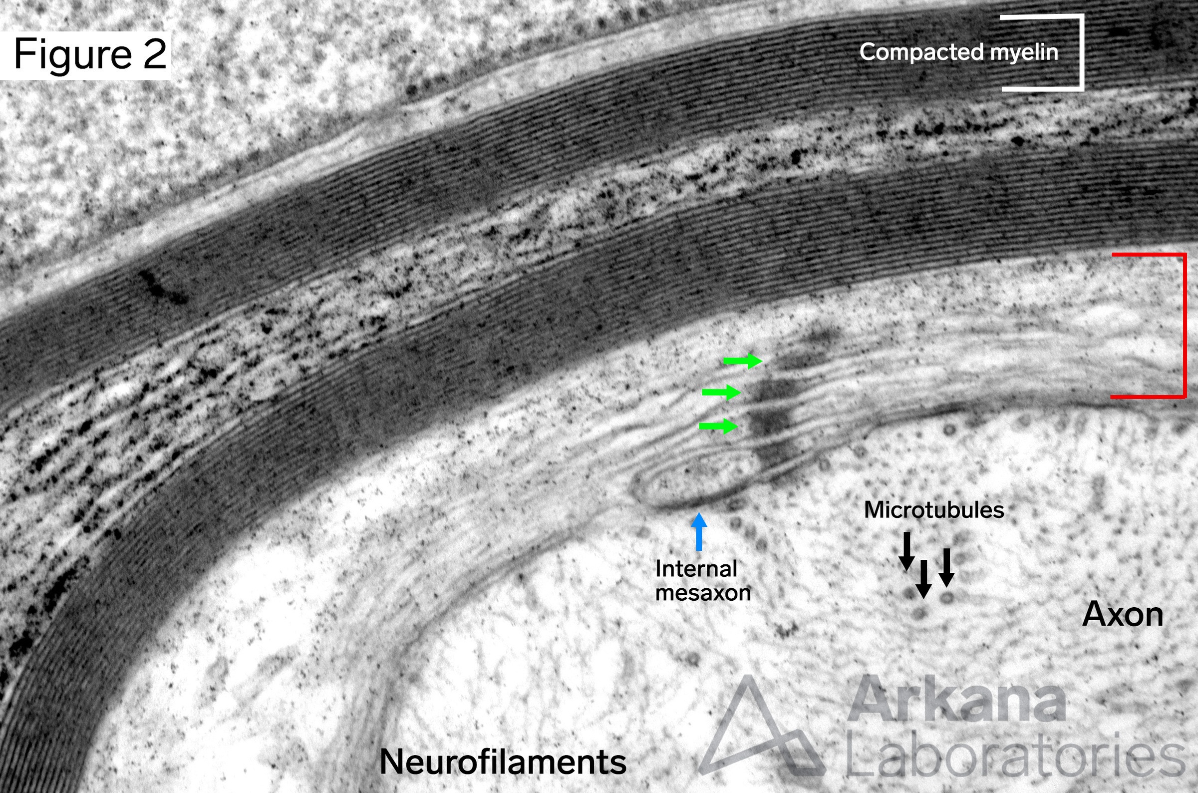
Myelin incisure (Schmidt-Lanterman incisure): channel-like region extending from inner to outer myelin sheath where Schwann cell cytoplasm is still present between Schwann cell membranes. Note: internal mesaxon and autotypic junctions (“desmosome stack”)
Myelin incisure (Schmidt-Lanterman incisure) in longitudinal orientation
Answer: Myelin incisure
The images show a myelinated axon that has been sectioned through the level of a myelin incisure (Schmidt-Lanterman incisure). These structures form a cytoplasmic channel from the innermost to outermost layers of the myelin sheath which is formed by successive multilayer wrapping of an individual peripheral nerve by a Schwann cell.
Despite being described some time ago, the precise function of the myelin incisure is not completely known. Possible functions include serving as a cytoplasmic channel connecting the inner and outermost aspects of the myelin sheath, formation and structural stability of myelin, regulation of adhesion, and signal transduction.
Why were the other answers incorrect?
Outer mesaxon: terminal end of outmost Schwann cell wrap (think outer edge of a tortilla rolled upon itself).
Cajal bands: cytoplasmic channels of Schwann cell cytoplasm which run between areas where the myelin sheath adheres to the overlying Schwann cell plasma membrane.
Axoplasm: cytoplasm of the axon.
References/Additional Reading
Weis J, Brandner S, Lammens M, Sommer C, Vallat JM. Processing of nerve biopsies: a practical guide for neuropathologists. Clin Neuropathol. 2012 Jan-Feb;31(1):7-23. doi: 10.5414/np300468. PMID: 22192700; PMCID: PMC3663462.
Quick note: This post is to be used for informational purposes only and does not constitute medical or health advice. Each person should consult their own doctor with respect to matters referenced. Arkana Laboratories assumes no liability for actions taken in reliance upon the information contained herein.