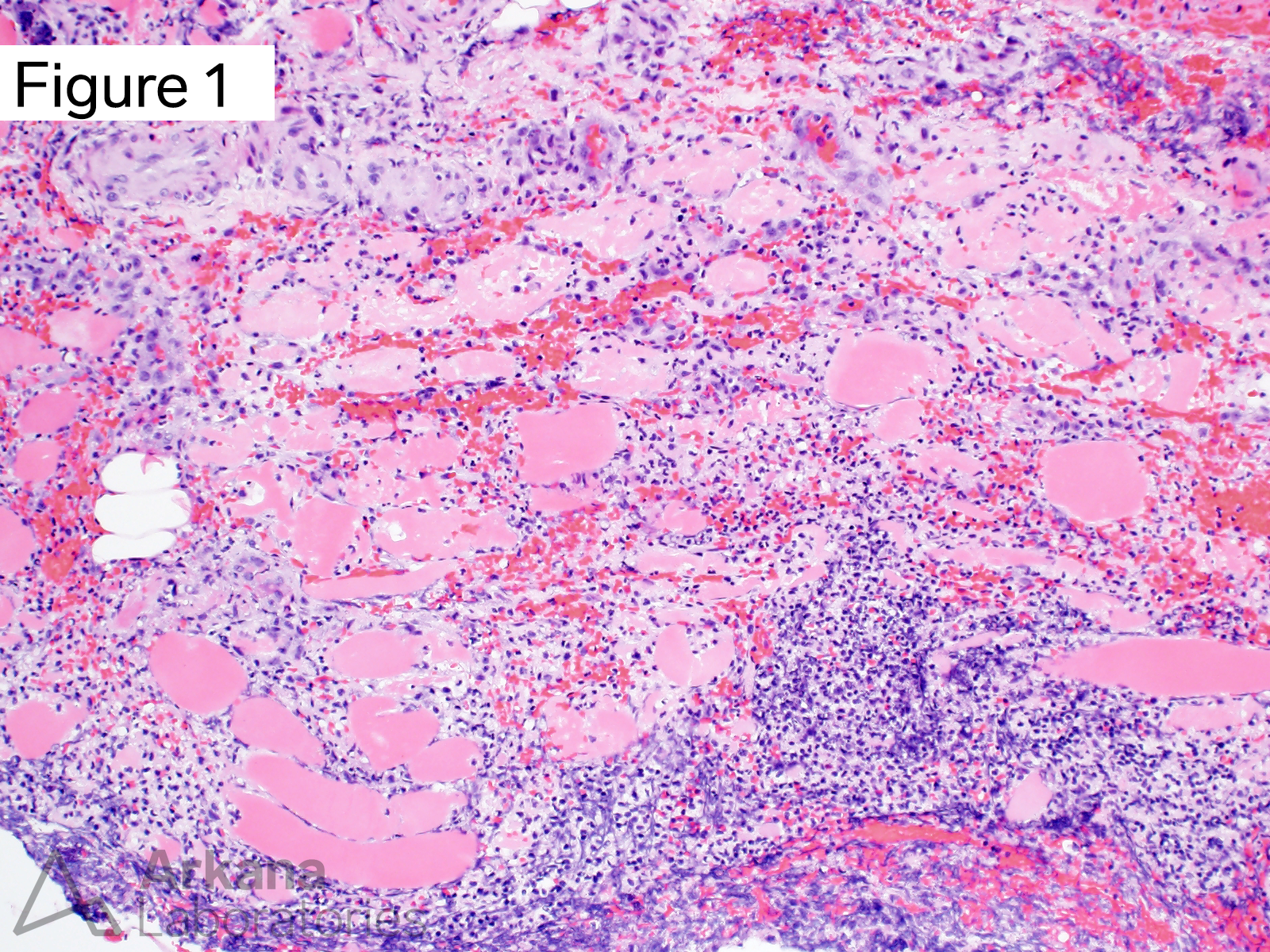
This 50-year-old female presents with progressively worsening swelling and pain involving her left hip and buttock of four days duration. CPK levels are in the 900 to 1200 range [normal 20-200 IU/L]. A muscle biopsy was performed.
A. Esterase enzyme histochemical stain
B. Grocott Methenamine Silver stain (GMS)
C. Tissue Gram stain
D. Modified Gomori Trichrome stain
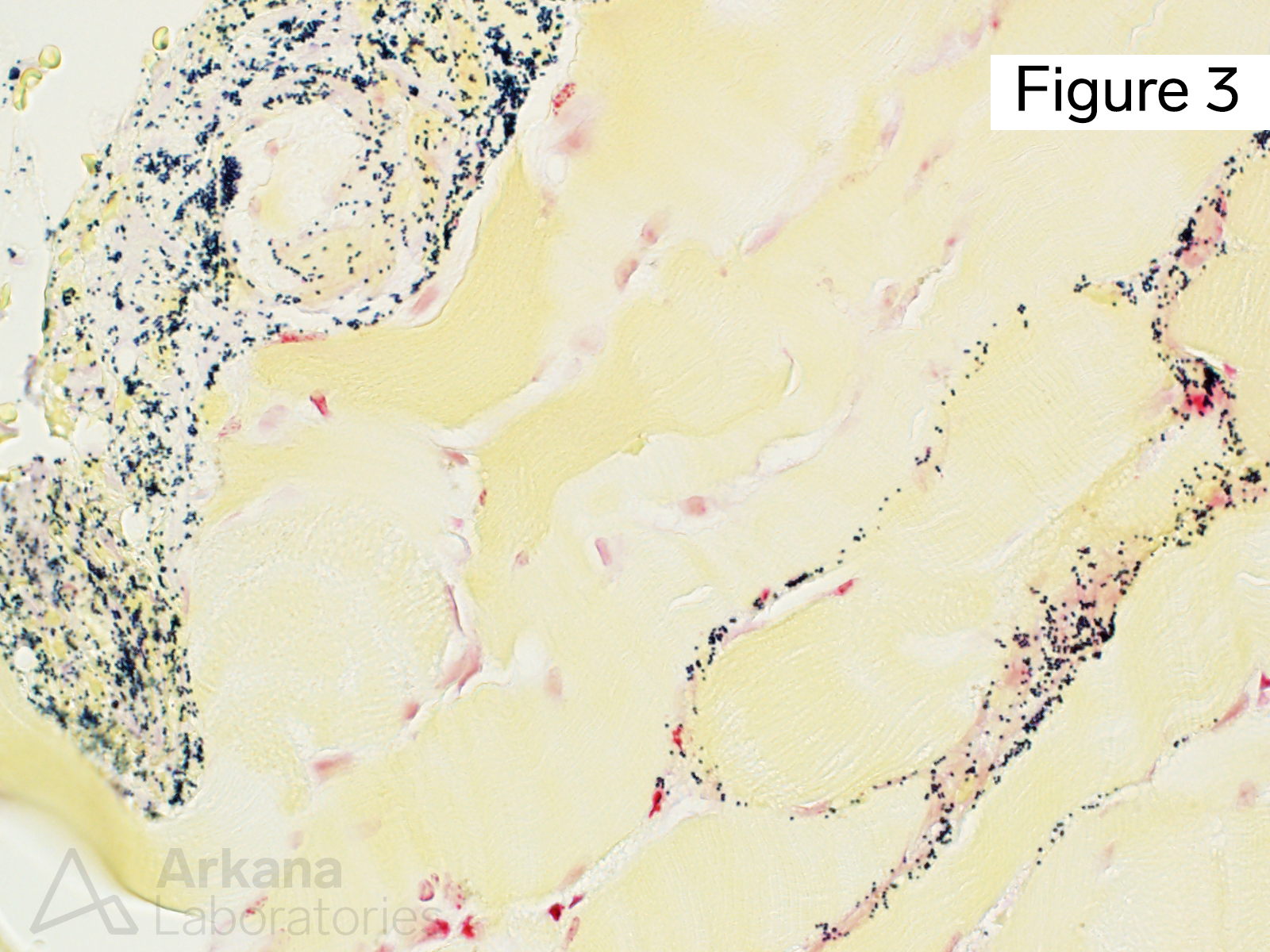
Answer: Tissue Gram stain
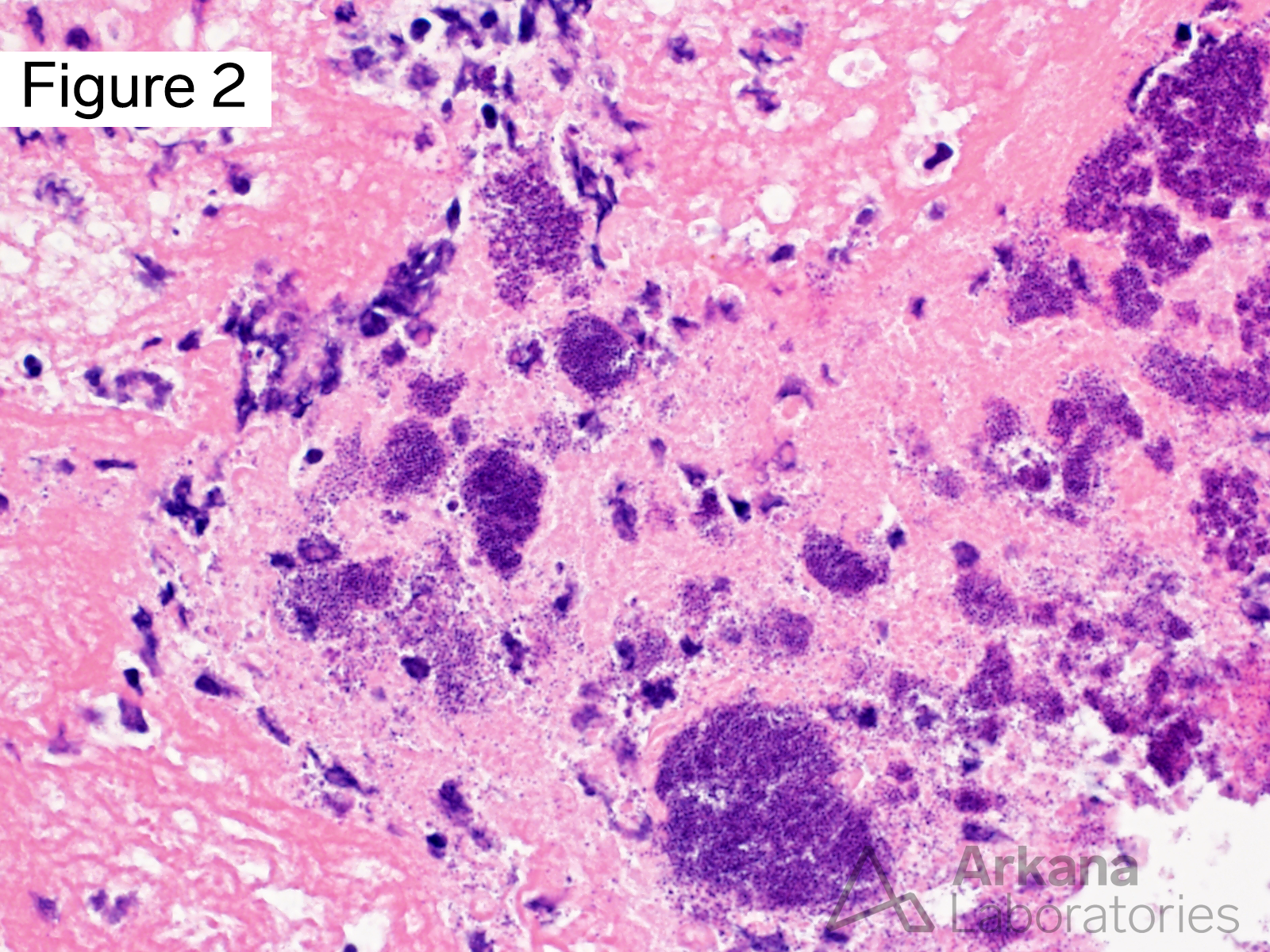
Figure 1 demonstrates inflamed skeletal muscle; Figure 2 shows bacterial cocci in clusters.
Tissue Gram stain is useful for attempting to further characterize the bacterial organisms, and demonstrates numerous Gram positive cocci (Image 3).
Why were the other answers wrong?
Esterase enzyme histochemical stain highlights denervated muscle fibers, neuromuscular junctions, and chronic inflammatory cells, but is not of use in evaluating infectious organisms.
Grocott methanamine silver stain is utility in evaluating fungal infections in surgical pathology and cytology specimens.
Modified Gomori Trichrome is routinely used in muscle biopsies, but in regards to bacterial infection does not provide additional information beyond what has already been seen on H&E stained sections
Quick note: This post is to be used for informational purposes only and does not constitute medical or health advice. Each person should consult their own doctor with respect to matters referenced. Arkana Laboratories assumes no liability for actions taken in reliance upon the information contained herein.