This 57-year-old patient presented with a left foot drop of several weeks duration. Laboratory studies showed elevated ESR, and negative/normal CRP, ANA, SSA, SSB, and ANCA. The patient was treated with steroids prior to nerve biopsy and reported subjective improvement in their strength.
What is the nature of the structures in Figure #2 indicated by arrows?
A. Artifact
B. Degenerating axons
C. Tomacula
D. Onion bulbs
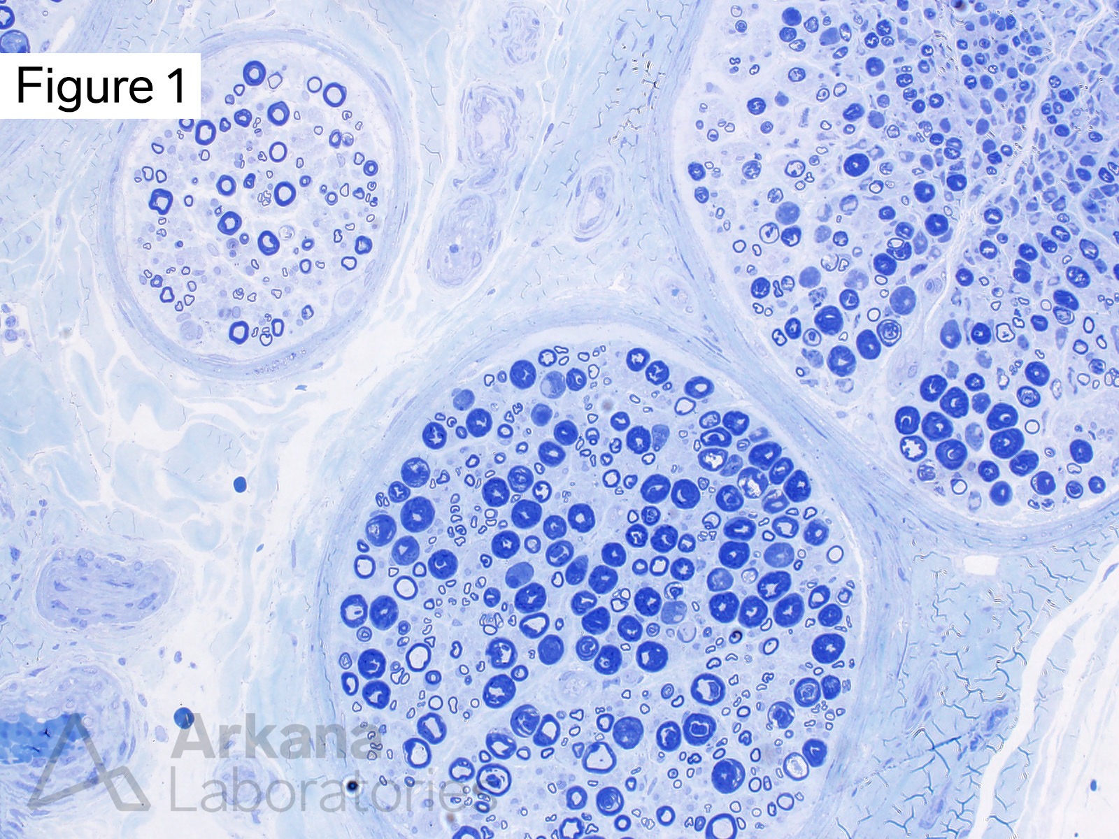
Medium magnification view showing three nerve fascicles. Lower magnification views such as this provide an overview, and context, for evaluation at higher magnification. In this case, note that the areas with thick appearing myelin in the two larger nerve fascicles have a patchy distribution. The smaller nerve fascicle at the upper left does not demonstrate this alteration.
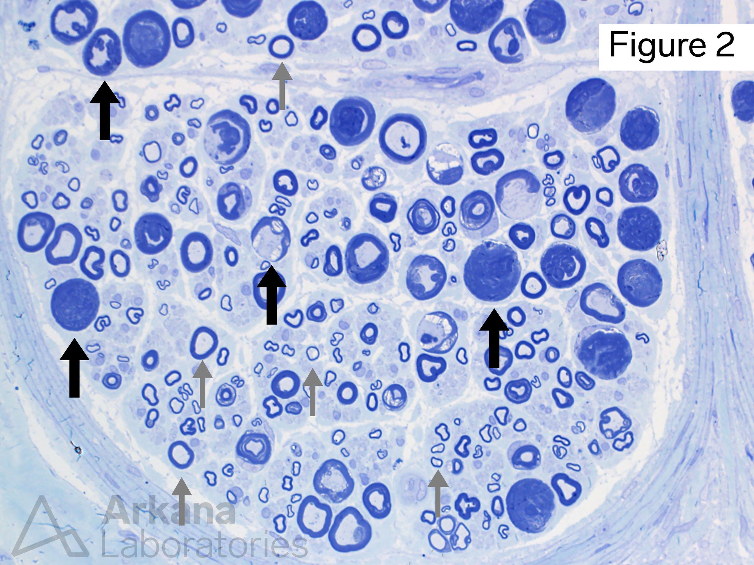
Higher magnification of one fascicle shows multiple large-diameter myelinated axons with irregularly thickened, lamellated, or cannon-ball-like appearance (thick arrows). These alterations represent partial traction or crush artifact. You can imagine that the lipid-rich myelin sheath will have a somewhat gelatinous consistency….. crushing or pulling on the nerve biopsy specimen can artifactually distort the appearance of the myelin sheath…. like squeezing toothpaste. Large diameter axons have more myelin and therefore are more susceptible to this artifact. Note the areas with relatively preserved appearing large diameter and small diameter myelinated axons (thin arrows). Lower magnification views such as this provide an overview, and context, for evaluation at higher magnification. In this case, note that the areas with thick appearing myelin in the two larger nerve fascicles have a patchy distribution. The smaller nerve fascicle at the upper left does not demonstrate this alteration.
Answer: Artifact
These changes represent partial crush and/or traction artifact. These alterations may mimic degenerating axons or tomacula. “Onion bulbs” represent redundant wraps of Schwann cells around a myelinated axon caused by multiple rounds (i.e. repetitive cycles) of demyelination and remyelination, and do not have this appearance.
References/Additional Reading
Mathis S, Magy L, Le Masson G, Richard L, Soulages A, Solé G, Duval F, Ghorab K, Vallat JM, Duchesne M. Value of nerve biopsy in the management of peripheral neuropathies. Expert Rev Neurother. 2018 Jul;18(7):589-602. doi: 10.1080/14737175.2018.1489240. Epub 2018 Jun 25. PMID: 29923431.
Chkheidze R, Pytel P. What Every Neuropathologist Needs to Know: Peripheral Nerve Biopsy. J Neuropathol Exp Neurol. 2020 Apr 1;79(4):355-364. doi: 10.1093/jnen/nlaa012. PMID: 32167544.
Quick note: This post is to be used for informational purposes only and does not constitute medical or health advice. Each person should consult their own doctor with respect to matters referenced. Arkana Laboratories assumes no liability for actions taken in reliance upon the information contained herein.