This 60-year-old patient presented with acute onset unilateral stroke-type symptoms. Imaging studies (brain CT and MRI) identified a left 5.0 cm frontal lobe hemorrhage with surrounding contrast enhancement and associated mass effect. CT scans of the chest, abdomen, and pelvis were negative for malignancy.
Microscopic examination of stereotactic biopsy tissue showed an infiltrative cellular neoplasm with necrosis. Based on additional findings seen in figures #1 – #6, what is your diagnosis? What is unusual about this particular neoplasm’s cytomorphology?
A. Metastatic carcinoma
B. Metastatic melanoma
C. Glioblastoma
D. Alexander disease

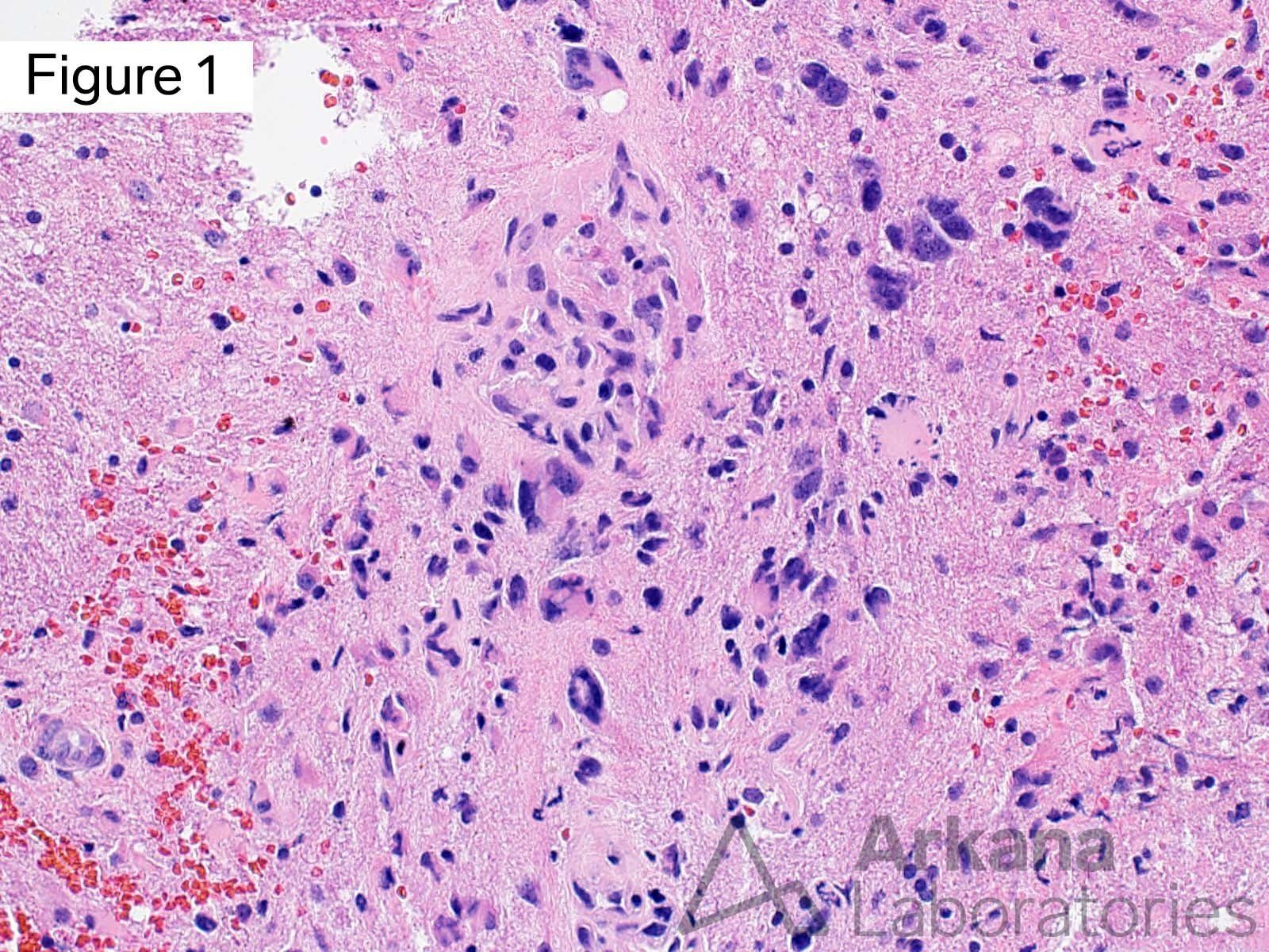
This image shows an area of viable neoplasm characterized by atypical appearing cells with astrocytic cytomorphology, and enlarged irregular hyperchromatic nuclei. Note the adjacent areas of necrosis.
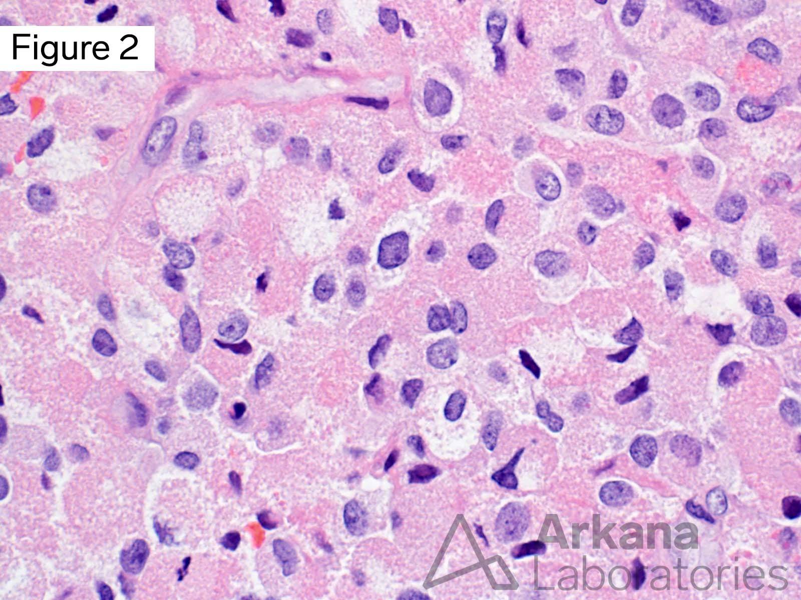
This image shows an area of relatively bland appearing neoplastic cells with oval to round nuclei, small nucleoli, and moderate amounts of cytoplasm with visible cytoplasmic borders. Such areas could be mistaken for macrophages.
Other areas showed more cytologic atypia and frequent mitotic figures.
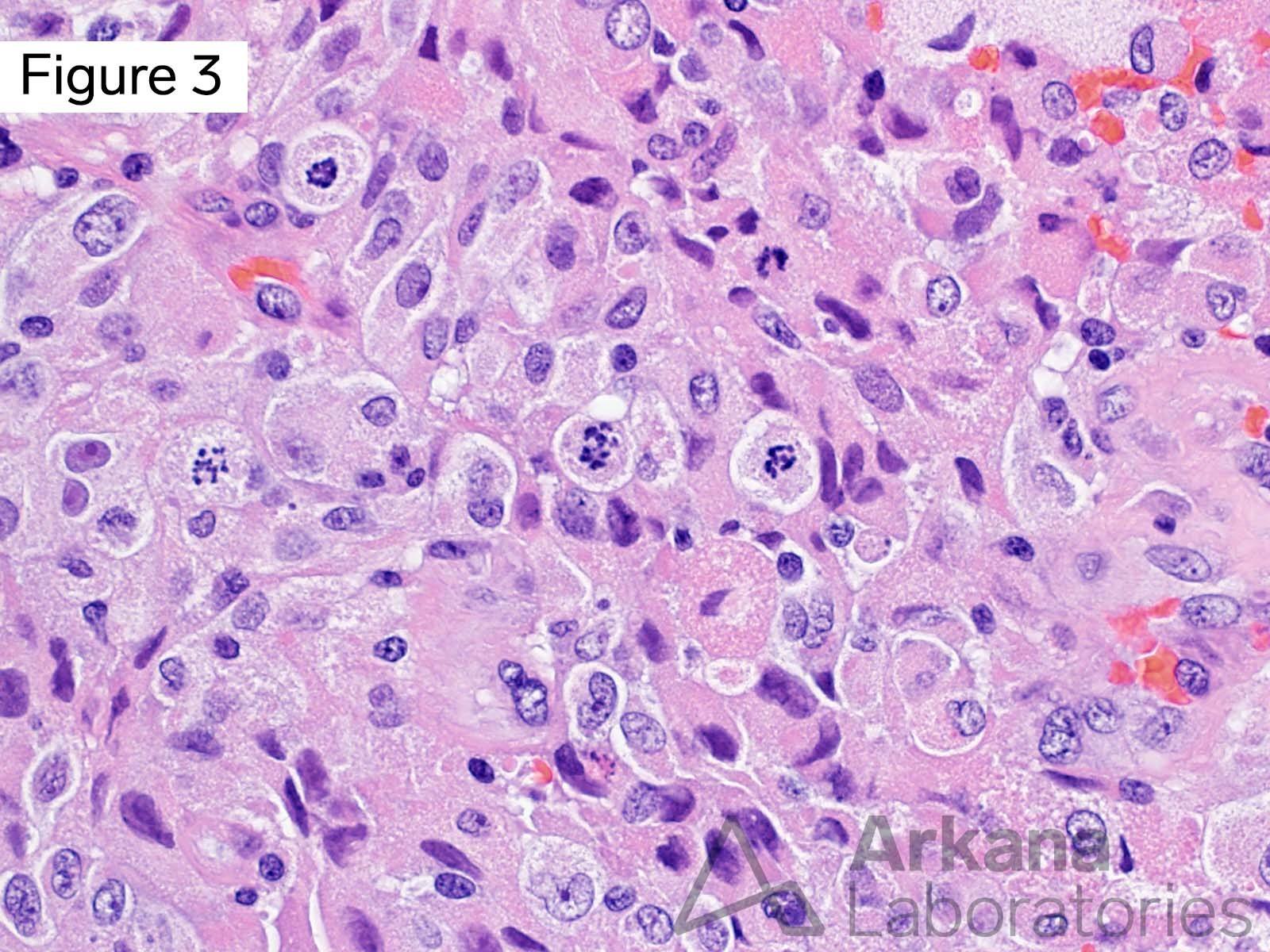
Many of the neoplastic cells showed variable amounts of somewhat refractile eosinophilic granular cytoplasmic material.
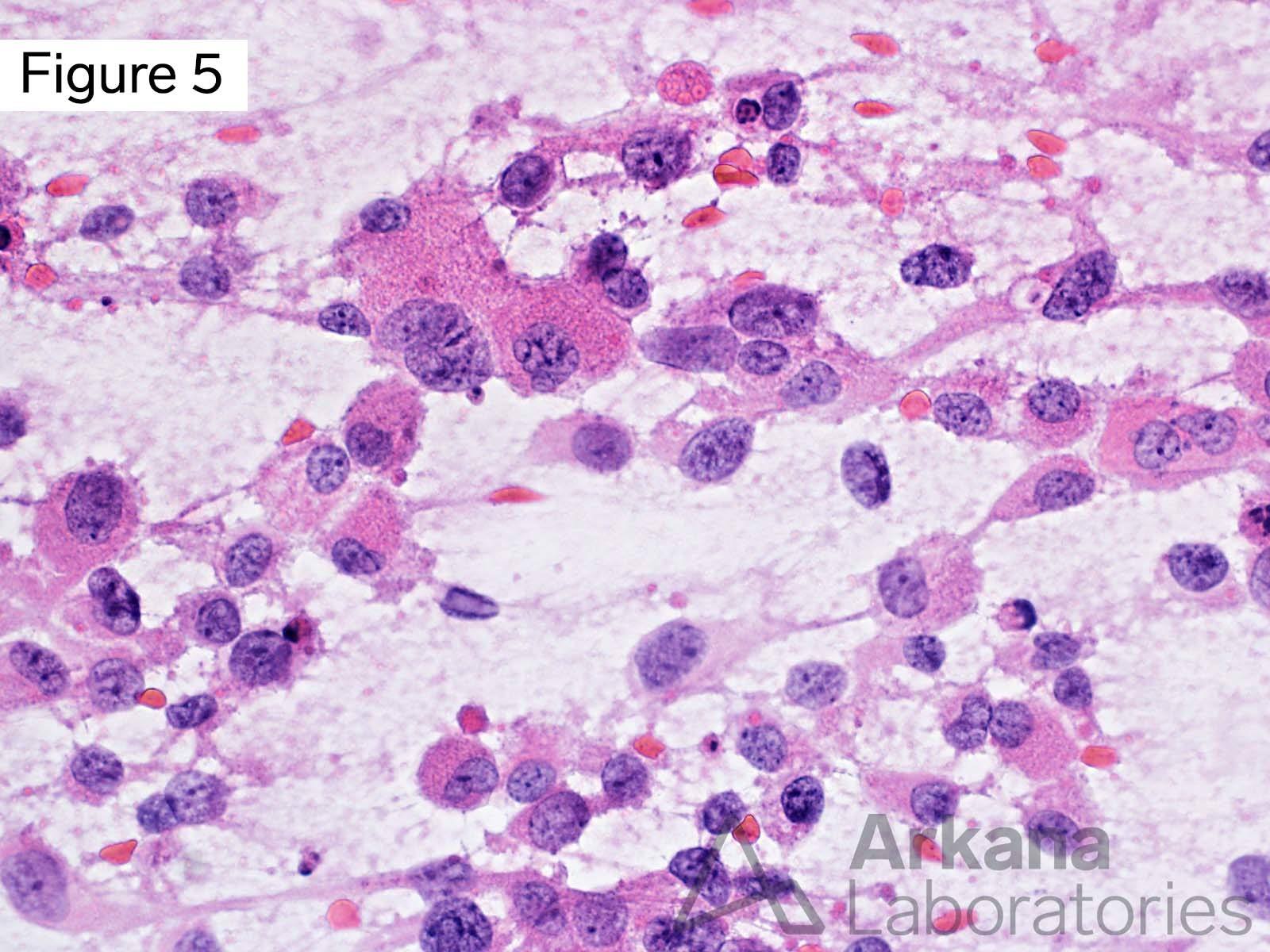
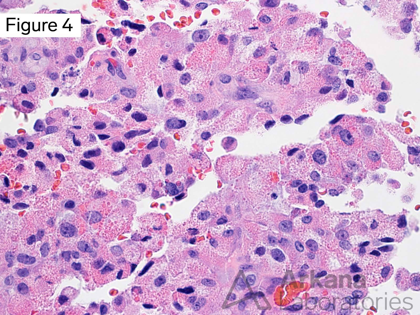
An intraoperative cytology preparation (“squash prep”) nicely demonstrates the eosinophilic granular cytoplasmic material.
Answer: Glioblastoma
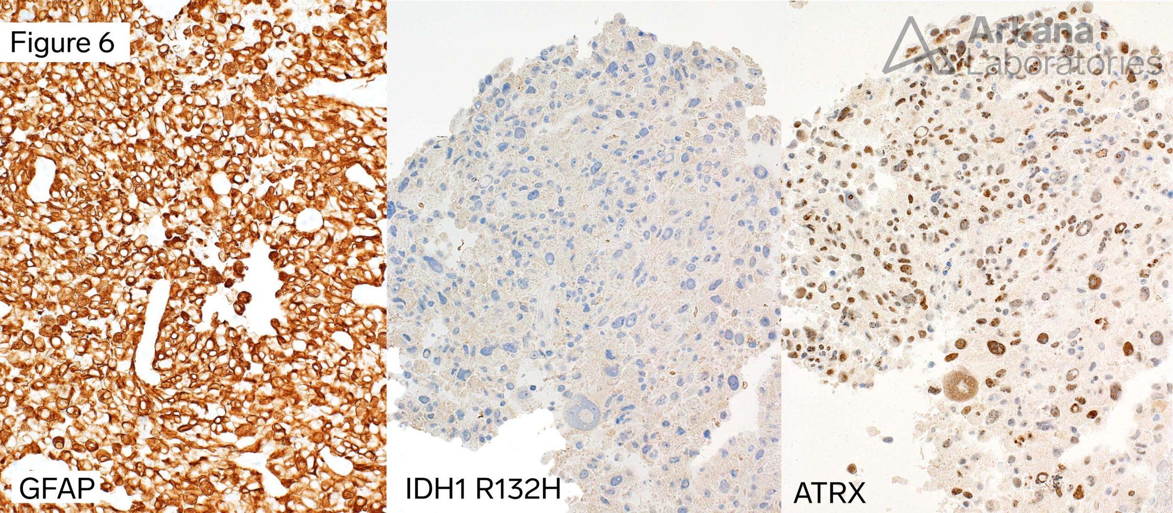
The images demonstrate the presence of a cellular GFAP-positive glial neoplasm with astrocytic cytomorphology. The presence of necrosis and brisk mitotic activity indicates that this is a high grade neoplasm. In a patient of this age, absence of immunohistochemical staining for IDH1 R132H mutant protein and retention of nuclear staining for ATRX, indicates that this is most consistent with the presence of an IDH-wildtype glioblastoma.
This lesional cells are unusual in showing many areas with histiocytoid to epithelioid cytomorphology and granular cytoplasm (“granular cell features”). Granular cell astrocytomas are uncommon and typically behave in an aggressive fashion. This aggressive behavior can be explained by a recently published study showing that granular cell astrocytomas (WHO grades 2 through 4) have molecular features of primary glioblastoma (please see references / additional reading below).
References/Additional Reading
Vizcaino MA, Palsgrove DN, Yuan M, Giannini C, Cabrera-Aldana EE, Pallavajjala A, Burger PC, Rodriguez FJ. Granular cell astrocytoma: an aggressive IDH-wildtype diffuse glioma with molecular genetic features of primary glioblastoma. Brain Pathol. 2019 Mar;29(2):193-204. doi: 10.1111/bpa.12657. Epub 2018 Oct 10. PMID: 30222900; PMCID: PMC6397086.
Schittenhelm J, Psaras T. Glioblastoma with granular cell astrocytoma features: a case report and literature review. Clin Neuropathol. 2010 Sep-Oct;29(5):323-9. doi: 10.5414/npp29323. PMID: 20860896.
Brat DJ, Scheithauer BW, Medina-Flores R, Rosenblum MK, Burger PC. Infiltrative astrocytomas with granular cell features (granular cell astrocytomas): a study of histopathologic features, grading, and outcome. Am J Surg Pathol. 2002 Jun;26(6):750-7. doi: 10.1097/00000478-200206000-00008. PMID: 12023579.
Quick note: This post is to be used for informational purposes only and does not constitute medical or health advice. Each person should consult their own doctor with respect to matters referenced. Arkana Laboratories assumes no liability for actions taken in reliance upon the information contained herein.