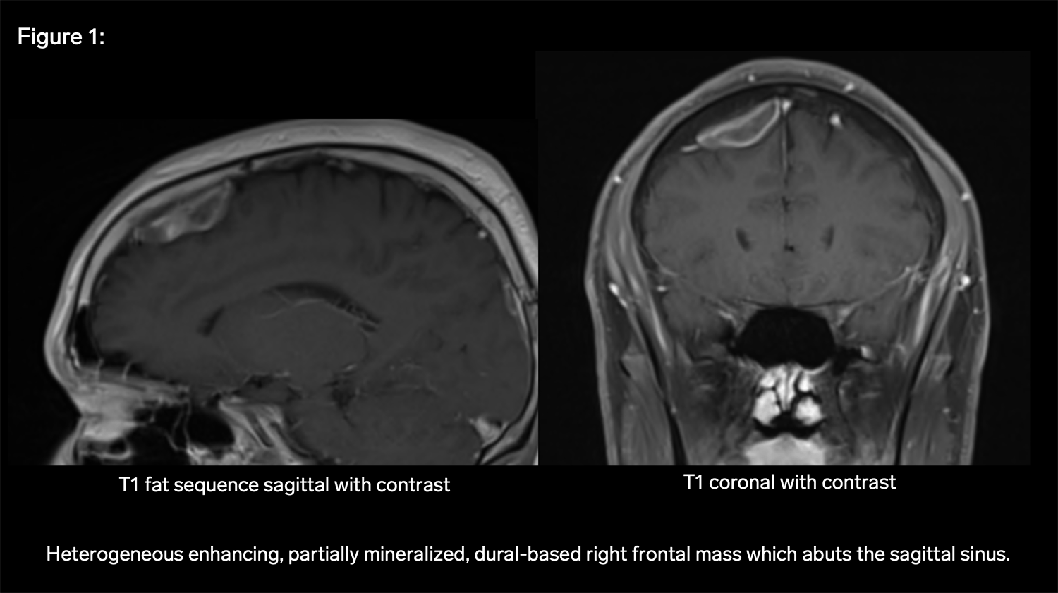
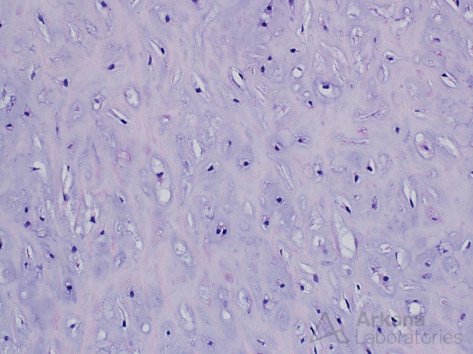
This 50-year-old patient presented with complaints of headaches. MRI showed an extra-axial parasagittal dural based mass which was gross totally resected. The lesional cells were negative for cytokeratin and EMA. The proliferative fraction as indicated by KI67 was very low.
What is your diagnosis based on Figures #1 and #2?
A. Chondroma
B. Meningioma
C. Glioma
D. Arachnoid cap cells

Answer:
The morphologic alterations are consistent with the diagnosis of chondroma. This is a rare lesion in this location.
References:
- Sullivan JC, Goldsmith J, Rojas R, Varma H, Kasper EM. Intracranial Dural Parafalcine Chondroma: Case Report and Systematic Review of the Literature. World Neurosurg. 2019 Feb;122:1-7. doi: 10.1016/j.wneu.2018.09.169. Epub 2018 Sep 29. PMID: 30273721.
- Chua MMJ, Bazarek SF, Meredith DM, Hsu L, Saris SC. Falcine chondroma: illustrative case. J Neurosurg Case Lessons. 2021 Feb 8;1(6):CASE20124. doi: 10.3171/CASE20124. PMID: 36045938; PMCID: PMC9394178.
Quick note: This post is to be used for informational purposes only and does not constitute medical or health advice. Each person should consult their own doctor with respect to matters referenced. Arkana Laboratories assumes no liability for actions taken in reliance upon the information contained herein.