Clinical History:
This 65-year-old patient presented with a two-month history of progressive proximal upper and lower extremity muscle weakness, and photosensitive skin rash involving their cheeks and lower neck. Their past medical history was significant for Rheumatoid arthritis. Home medications included prednisone and leflunomide. Muscle biopsy was performed to evaluate for a myopathy.
Question #1: What is your diagnosis?
A. Polymyositis
B. Vasculitis
C. Dermatomyositis
D. Sarcoidosis
Question #2: What feature does the H&E visualized under fluorescence microscopy nicely demonstrate?
A. Disrupted elastic lamina
B. Normal elastic lamina
C. Redundant elastic lamina
D. Amyloid
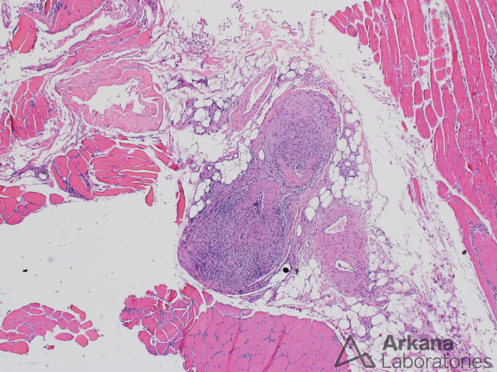
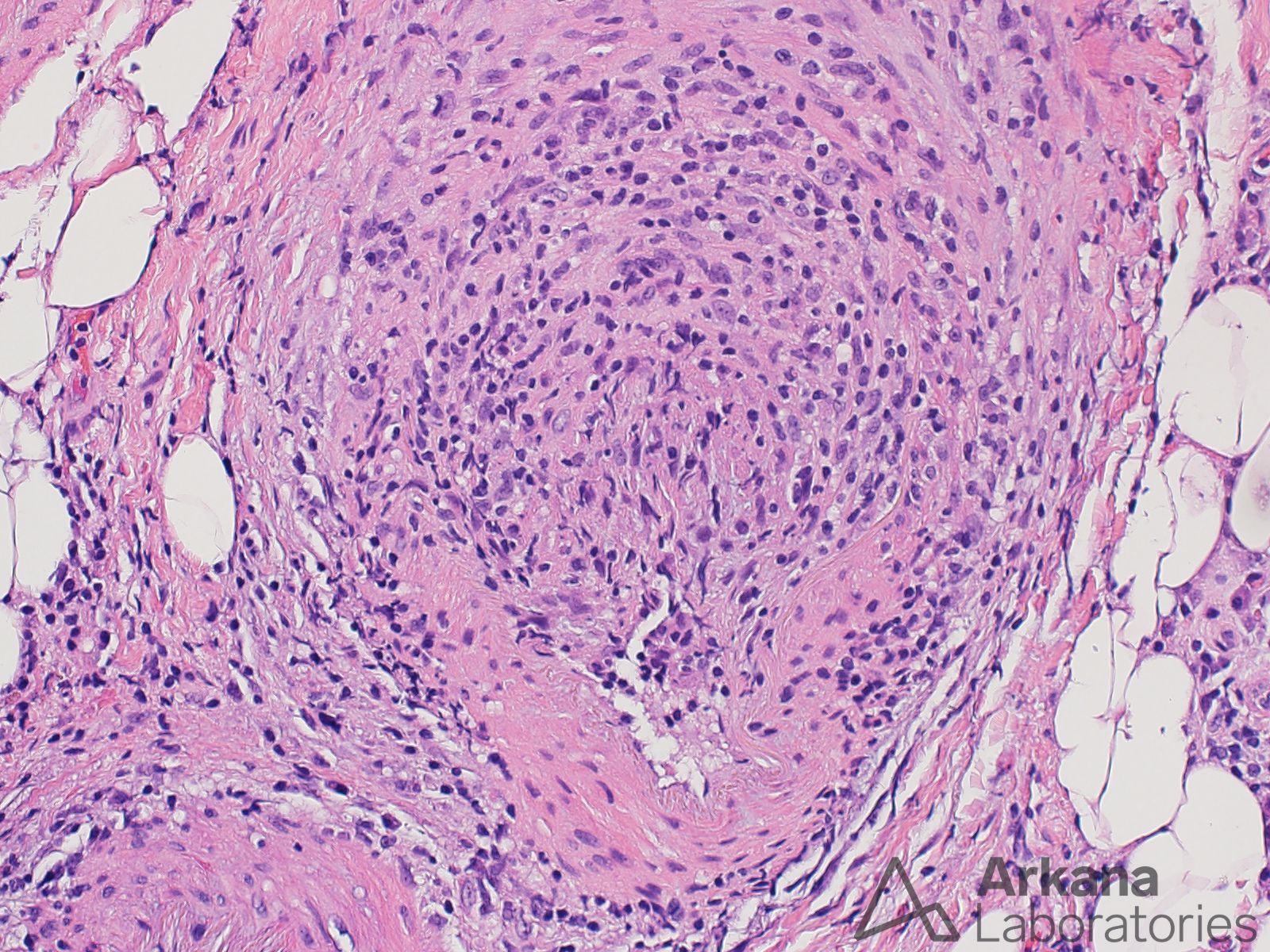
Answer #1: Vasculitis
The H&E stain images demonstrate two blood vessels surrounded by chronic inflammatory cells and macrophages that also infiltrate the blood vessel walls, and areas of fibrinoid necrosis. The overall morphologic abnormalities are consistent with the presence of a small vessel vasculitis. In the context of this patient’s clinical history, vasculitis may be seen in the setting of Rheumatoid arthritis (rheumatoid vasculitis) and may also rarely be induced by leflunomide (i.e. drug induced vasculitis).
The features provided in the images are not those of polymyositis or dermatomyositis. No well-formed granulomata characteristic of sarcoidosis are seen. However, the granulomata of sarcoidosis may involve blood vessels (so-called sarcoid vasculopathy).
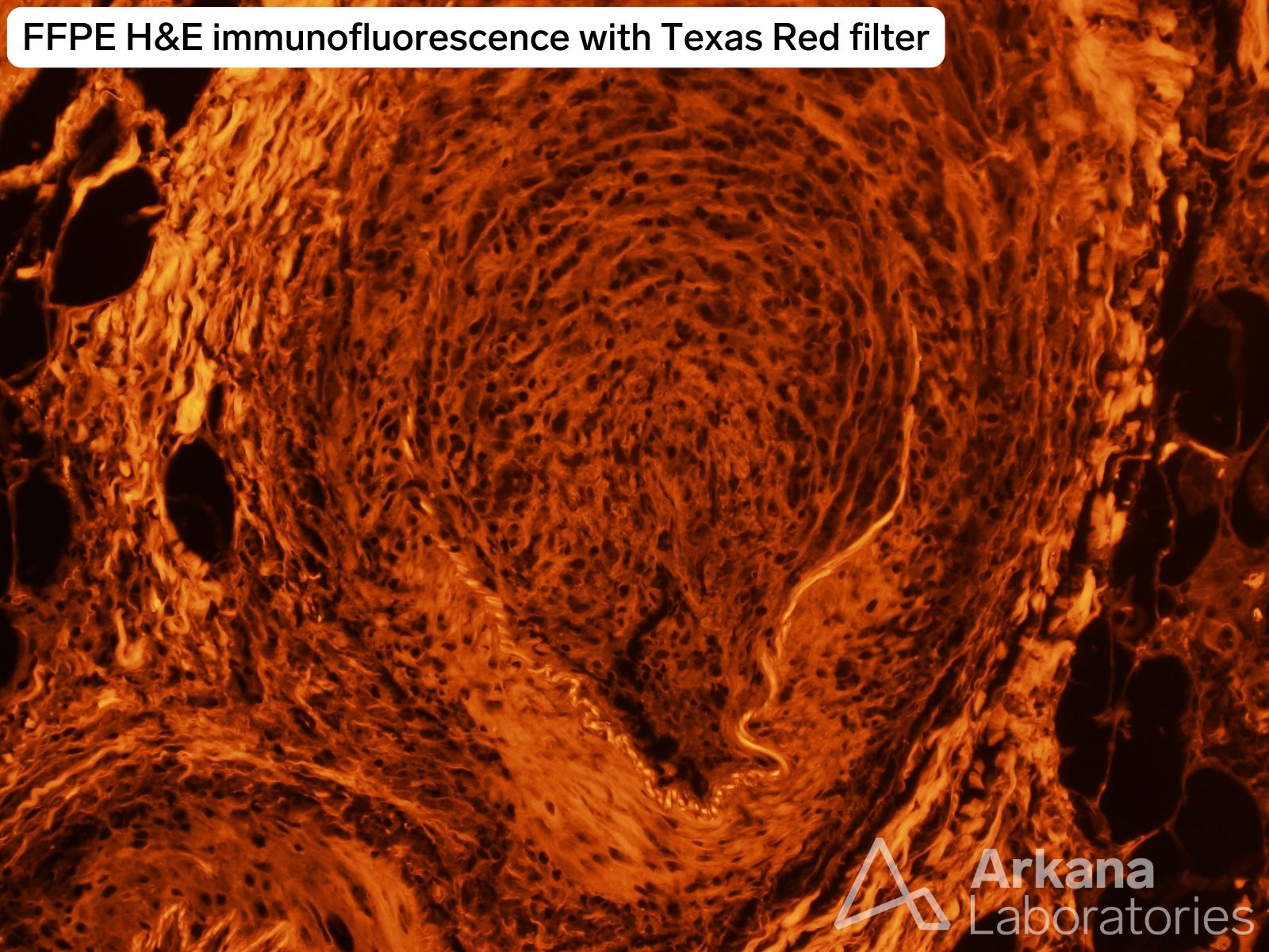
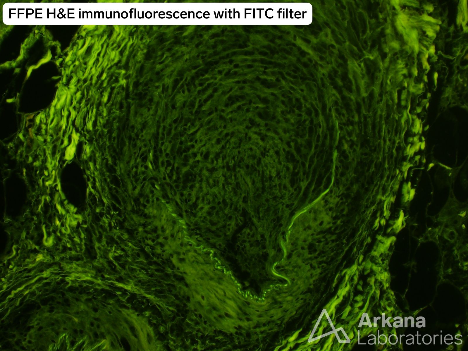
Answer #2: Disrupted elastic lamina
Eosin, present in the H&E stain (hematoxylin-eosin), is autofluorescent. Because the elastic lamina in arteries has an enhanced affinity for eosin it may be visualized on fluorescence microscopy on an H&E-stained section. In some instances it can serve as an alternative to an elastin stain.
The elastic lamina is not intact appearing or redundant appearing. Amyloid is best demonstrated with Congo Red stain, and is typically not highlighted on an H&E.
Reference(s) / Additional Reading:
- de Carvalho HF, Taboga SR. Fluorescence and confocal laser scanning microscopy imaging of elastic fibers in hematoxylin-eosin stained sections. Histochem Cell Biol. 1996 Dec;106(6):587-92. doi: 10.1007/BF02473274. PMID: 8985747.
- Makol A, Crowson CS, Wetter DA, Sokumbi O, Matteson EL, Warrington KJ. Vasculitis associated with rheumatoid arthritis: a case-control study. Rheumatology (Oxford). 2014 May;53(5):890-9. doi: 10.1093/rheumatology/ket475. Epub 2014 Jan 17. PMID: 24441152; PMCID: PMC3999374.
- Mertz P, Wollenschlaeger C, Chasset F, Dima A, Arnaud L. Rheumatoid vasculitis in 2023: Changes and challenges since the biologics era. Autoimmun Rev. 2023 Sep;22(9):103391. doi: 10.1016/j.autrev.2023.103391. Epub 2023 Jul 17. PMID: 37468085.
- Macdonald J, Zhong T, Lazarescu A, Gan BS, Harth M. Vasculitis associated with the use of leflunomide. J Rheumatol. 2004 Oct;31(10):2076-8. PMID: 15468379.
Quick note: This post is to be used for informational purposes only and does not constitute medical or health advice. Each person should consult their own doctor with respect to matters referenced. Arkana Laboratories assumes no liability for actions taken in reliance upon the information contained herein.