

Clinical History:
This 40-year-old patient presented with progressive neuropathic symptoms affecting their distal lower extremities and hands. A sural nerve biopsy was performed.
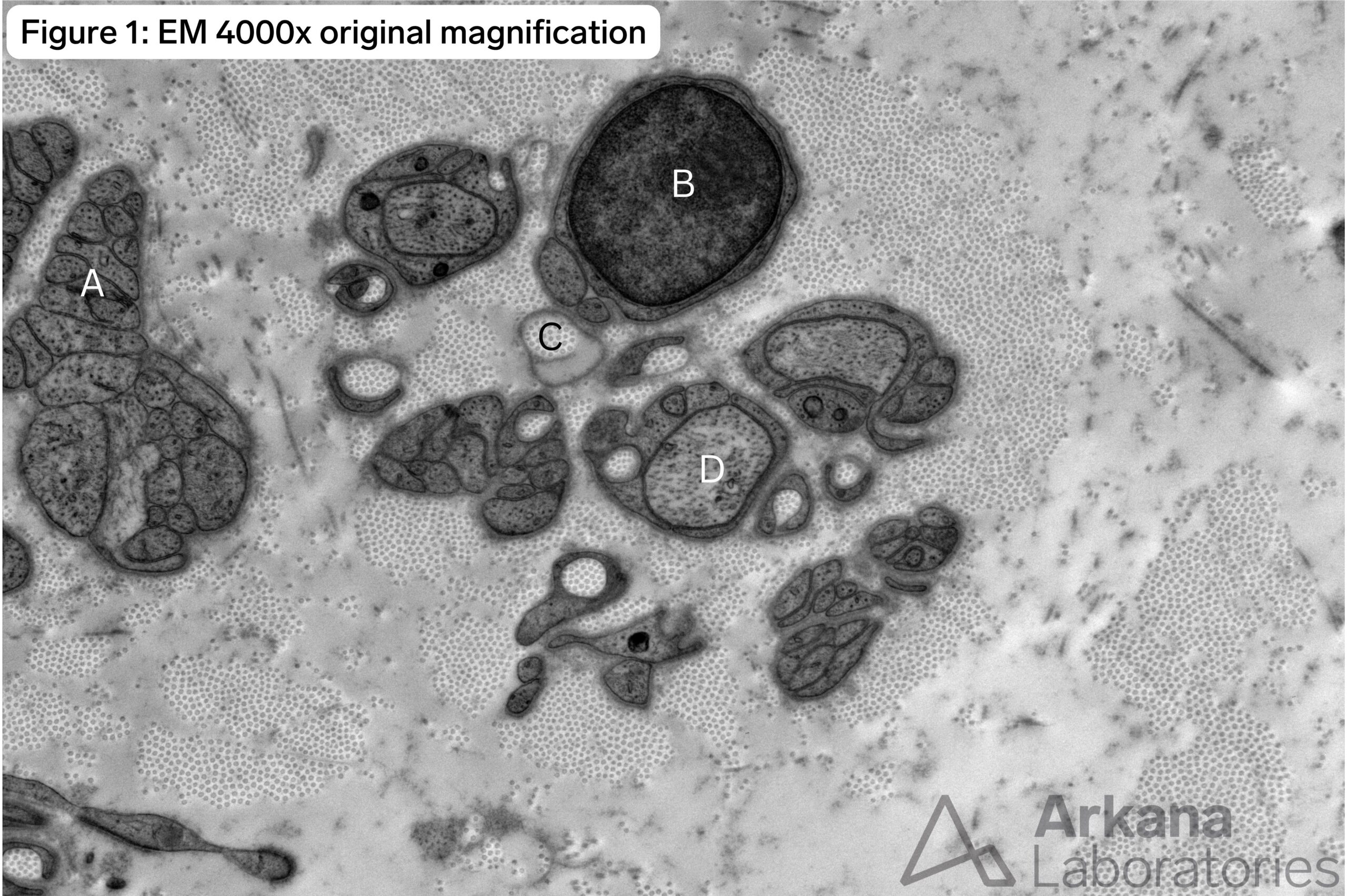
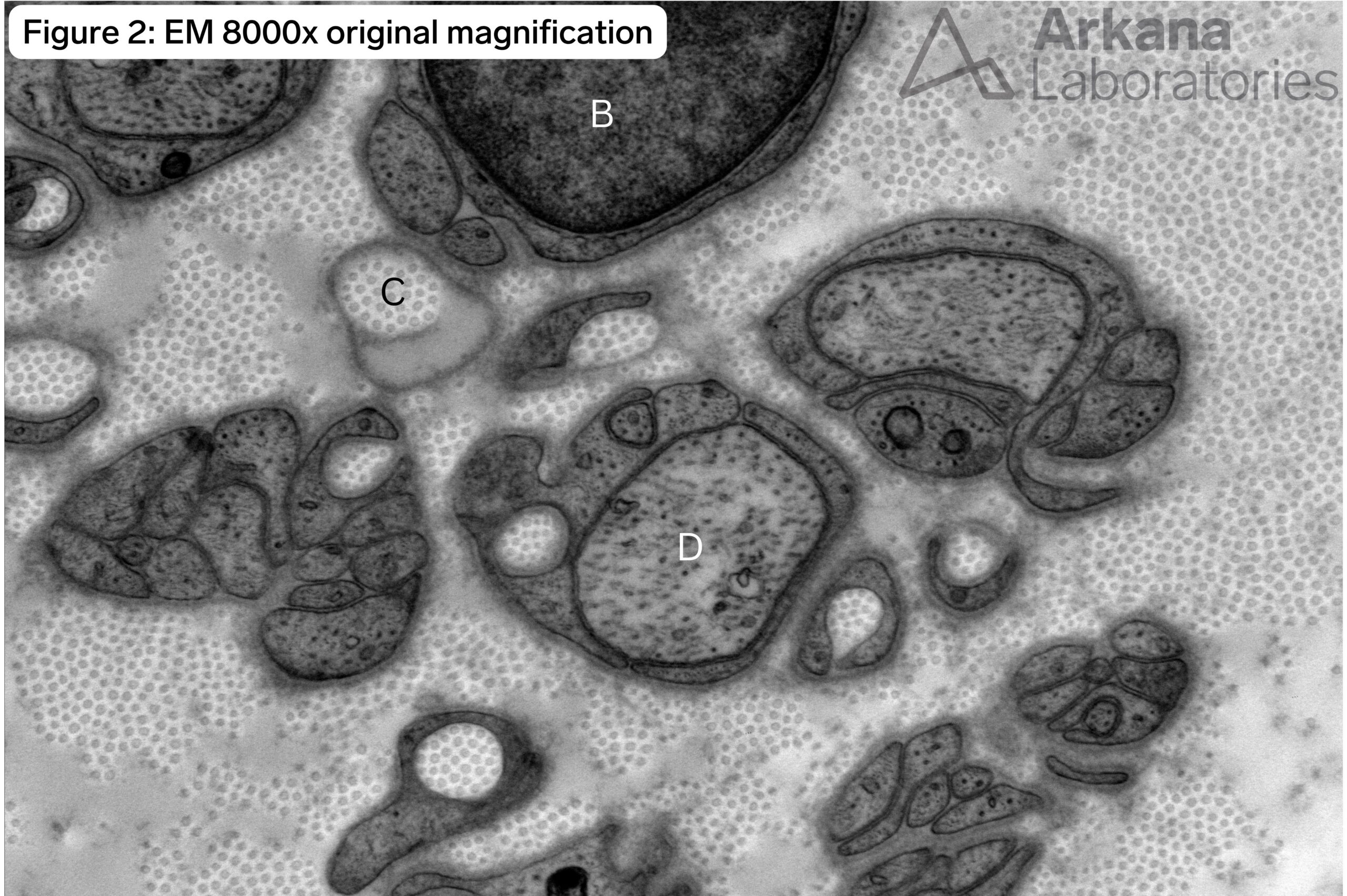
In the supplied medium and high magnification images from electron microscopy, which of the labeled structures represents an intact unmyelinated small diameter axon?
A.
B.
C.
D.
Answer:
D.
- The small diameter unmyelinated axon marked by “D” shows microtubules and intermediate filaments within its axoplasm. These are best seen on high magnification images.
- “A”, “B”, “C” and “D” are all part of a Schwann cell subunit. “A” is a denervated non-myelinating Schwann cell profile; “B” is the nucleus of a non-myelinating Schwann cell; “C” is a collagen pocket (see prior Arkana Neuronotes: https://www.arkanalabs.com/collagen-pockets-in-nerve-biopsy/)
Quick note: This post is to be used for informational purposes only and does not constitute medical or health advice. Each person should consult their own doctor with respect to matters referenced. Arkana Laboratories assumes no liability for actions taken in reliance upon the information contained herein.


