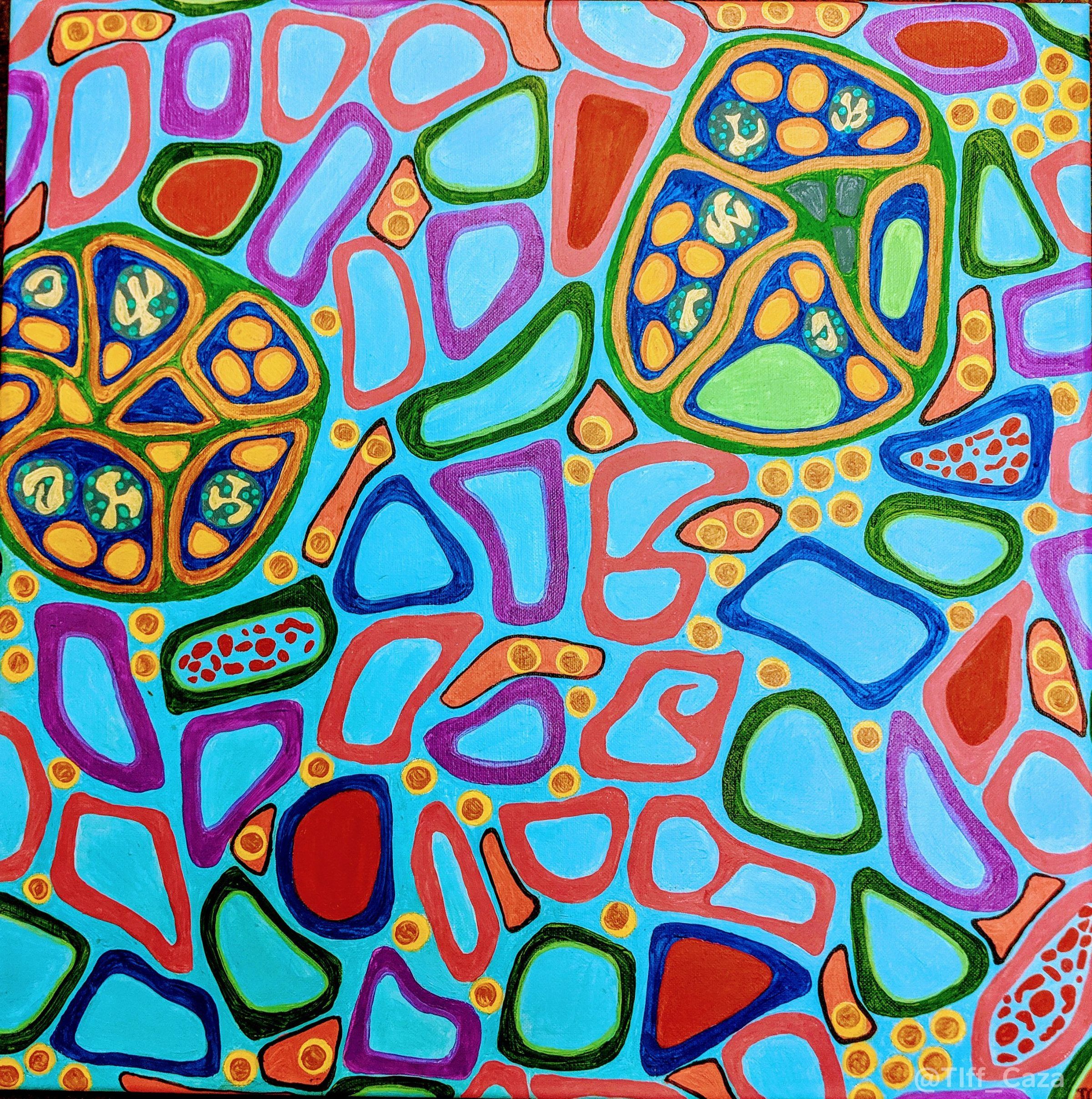
The above painting shows endocapillary hypercellularity with mononuclear cells and neutrophils, as well as hyaline deposits within glomeruli. Red blood cell casts are seen within focal tubular lumens, and the tubules are widely spaced due to interstitial edema and inflammation. These findings can be seen within acute proliferative glomerulonephritis. Other proliferative changes within glomeruli seen in acute proliferative glomerulonephritis include mesangial hypercellularity and crescent formation.
The differential diagnosis for proliferative glomerulonephritis is broad and includes infection-associated glomerulonephritis including post-streptococcal glomerulonephritis, focal or diffuse lupus nephritis, shunt nephritis, cryoglobulinemic glomerulonephritis, hepatitis-associated glomerulonephritis, and IgA nephropathy (or Henoch-Schonlein purpura nephritis). Other considerations include bacterial endocarditis (which is more commonly associated with crescentic glomerulonephritis), HIV infection, proliferative glomerulonephritis with monoclonal IgG deposits, C3 glomerulonephritis, myeloproliferative neoplasm-associated glomerulonephritis, and medications. Medications that can induce proliferative glomerulonephritis are those associated with drug-induced lupus and include procainamide, quinidine, hydralazine, isoniazid, minocycline, etanercept, and infliximab. In drug-induced lupus, patients typically develop antinuclear antibodies, anti-histone antibodies, and anti-ssDNA antibodies; while patients with systemic lupus erythematosus with nephritis commonly have anti-dsDNA and anti-Smith antibodies.
Immunofluorescence findings and clinical correlation with the patient’s history can be helpful to narrow this broad differential diagnosis. A “full house” immunofluorescence study with positivity for all three immunoglobulin heavy chains (IgA, IgG, and IgM) as well as complement components (C3 and C1q) can be seen in lupus nephritis, drug-induced lupus, and HIV infection (HIV-associated immune complex disease of the kidney, or HIVICK). IgA can be the dominant immune reactant in IgA nephropathy, Henoch-Schonlein purpura nephritis (also known as IgA vasculitis), and in IgA-dominant infection-associated glomerulonephritis. C3 can be the dominant immune reactant in infection-associated glomerulonephritis, shunt nephritis, and in C3 glomerulonephritis. IgG and IgM are the dominant immune complex reactants in cryoglobulinemic glomerulonephritis and be also expressed in infection-associated glomerulonephritis. In proliferative glomerulonephritis with monoclonal IgG deposits, light chain and IgG subclass restriction are seen.
Laboratory studies to aid in narrowing down the differential diagnosis of proliferative glomerulonephritis include serologic studies for ANA, anti-dsDNA, anti-histone antibodies, Hepatitis B, Hepatitis C, HIV, complement components (C3, C4, and CH50), and serum and urine protein electrophoresis. A thorough workup to exclude infections may also be required. Since proliferative glomerulonephritis can be a non-specific pattern of glomerular injury, clinicopathologic correlation may help identify a potential etiology, which is important as some patients may require systemic immunosuppression, while others would require treatment of an underlying infection or hematologic neoplasm.
Quick note: This post is to be used for informational purposes only and does not constitute medical or health advice. Each person should consult their own doctor with respect to matters referenced. Arkana Laboratories assumes no liability for actions taken in reliance upon the information contained herein.
