Search & Filter All Posts
All Articles
(8 Results)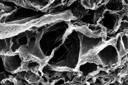
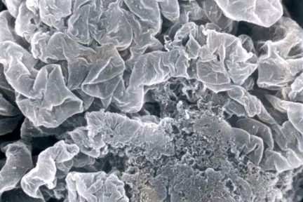
Fibrocellular Crescents with Smooth Basement Membrane Lining Lacunar Spaces
Its time for another eyeSCANdy image! This acellular scanning EM image shows fibrocellular crescents with smooth basement membrane-like material lining…
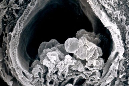
Glomerulus with Ischemic Collapse
Todays eyeSCANdy image shows acellular scanning EM showing a glomerulus with ischemic collapse. Photo credits to Dr. Stephen Bonsib.
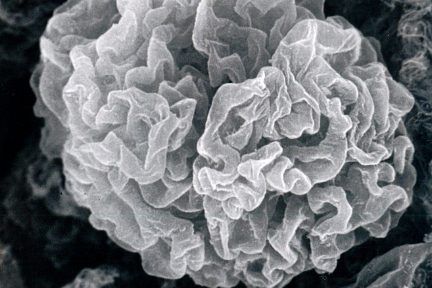
Subpodcocytic Capillary Loop Basement Membranes After Removal of Podocytes
This eyeSCANdyimage shows acellular scanning EM of a normal glomerular tuft showing the subpodcocytic capillary loop basement membranes following removal…

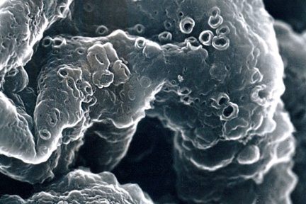
Glomerulus with a Membranoproliferative Pattern
This eyeSCANdy image shows acellular scanning EM showing glomerular basement membrane duplication at high power in a glomerulus with a…
Glomerular Capillary Loops with Advanced Membranous Glomerulonephritis
Todays eyeSCANdy image shows acellular scanning EM of glomerular capillary loops from a biopsy with advanced membranous glomerulonephritis stage IV…
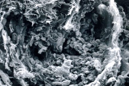
Acellular Scanning EM with Fibrocellular Crescent
This eyeSCANdy image shows acellular scanning EM with a fibrocellular crescent with entrapped RBCs. The glomerulus is not present.
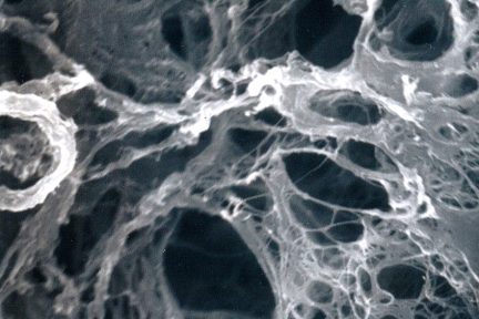
Acellular Scanning EM with Empty Lacunar Spaces
Todays eyeSCANdy image shows acellular scanning EM with empty lacunar spaces of a fibrocellular crescent previously occupied by cells.
Segmental Necrosis
Todays eyeSCANdy image shows acellular scanning EM with normal glomerular tuft at top and segmental necrosis at bottom.…