Search & Filter All Posts
Results for eyeSCANdy
(86 Results)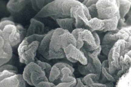
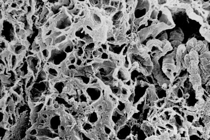
Membranous Glomerulonephritis
Todays eyeSCANdy image is of an acellular scanning EM of a glomerulus from a biopsy with membranous glomerulonephritis, stage II…
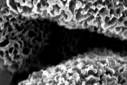
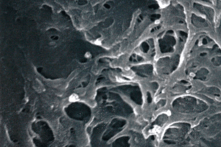
Membranous Glomerulonephritis Stage II
Todays acellular scanning EM from a biopsy with membranous glomerulonephritis, stage II shows a reticular pattern of the subepithelial GBM.…
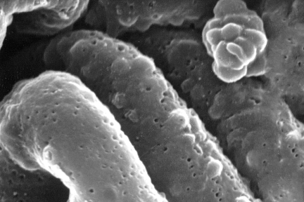
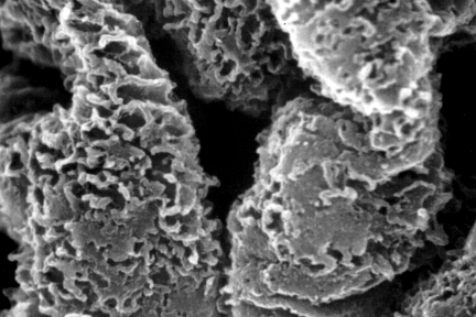
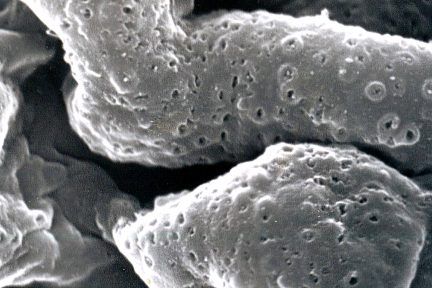
Membranous Glomerulonephritis Stage I
Todays eyeSCANdy cellular scanning EM image from a biopsy with membranous glomerulonephritis, stage I shows numerous small shallow depressions representing…

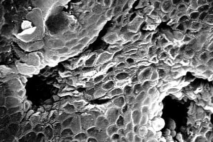
Glomerular Capillary Loops
This acellular scanning EM shows several glomerular capillary loops from a biopsy with membranous glomerulonephritis, stage II. The extracted deposits…
Squamoid-Appearing Surface Urothelial Cells
Todays eyeSCANdy scanning EM of normal papillary tip shows ostia of ducts of Bellini and squamoid-appearing surface urothelial cells. Photo…
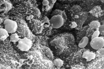
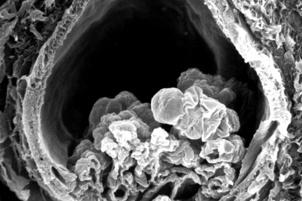
Red Blood Cells Within A Collecting Duct
Todays eyeSCANdy image shows red blood cells, some dysmorphic, within a collecting duct! Photo credits to Dr. Stephen Bonsib!
Macula Densa Basement Membrane Tunnels for Epithelial Cell Process to Contact Lacis Cells
Todays eyeSCANdy image shows macula densa basement membrane tunnels for epithelial cell process to contact lacis cells. Photo credits to…
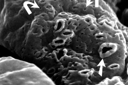
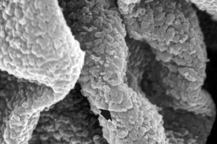
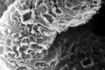
Segmental Membranous Glomerulonephritis
This acellular SEM of segmental membranous glomerulonephritis shows an admixture of craters (straight arrow) and tiny pit-like indentations (curved arrows)…
Depressions and Larger “Craters”
Todays acellular scanning EM from a biopsy with membranous glomerulonephritis shows depressions and larger “craters” along the subepithelial aspect of…
Subepithelial Immune Deposits Carpeting the GBM Surface
Todays eyeSCANdy image of an acellular scanning EM from a biopsy with membranous lupus nephritis shows subepithelial immune deposits (arrows,…
Immune Complexes
Todays acellular scanning EM is from a biopsy with membranous glomerulonephritis, stage I and shows numerous small shallow depressions representing…
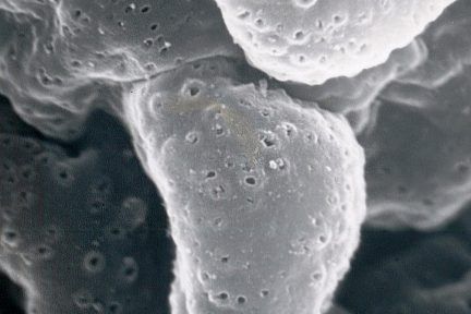
Extracted Immune Complexes
Todays acellular scanning EM from a biopsy with membranous glomerulonephritis, stage I shows numerous small shallow depressions representing the site…
Fibrocellular Crescent
This acellular scanning EM shows a fibrocellular crescent with a small remnant of the glomerular tuft (middle right). Photo credits…
Glomerular Basement Membrane With A Reticular Appearance And Occasional “Craters”
This acellular scanning EM from a biopsy with membranous lupus nephritis shows glomerular basement membrane (large arrow) with a reticular…
Glomerulus with Ischemic Collapse
Todays eyeSCANdy image shows acellular scanning EM showing a glomerulus with ischemic collapse. Photo credits to Dr. Stephen Bonsib!