Search & Filter All Posts
All Articles
(6 Results)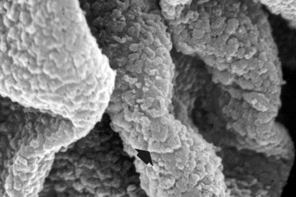
Subepithelial Immune Deposits Carpeting the GBM Surface
Todays eyeSCANdy image of an acellular scanning EM from a biopsy with membranous lupus nephritis shows subepithelial immune deposits (arrows,…
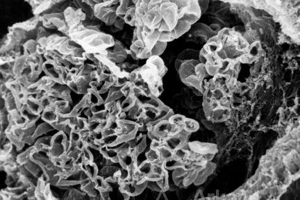
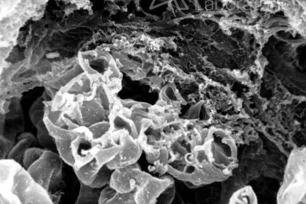
Glomerulus with Fibrocellular Crescent
Todays #eyesSCANdy image shows acellular scanning EM of a glomerulus with fibrocellular crescent (right). Photo credits to Dr. Stephen Bonsib!
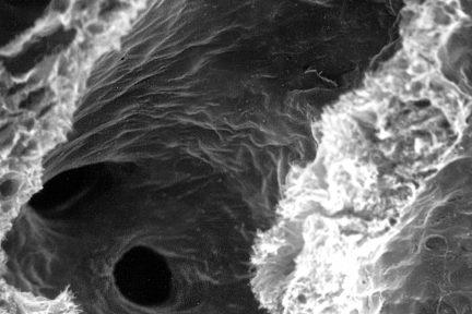
Ostia of Entering Venules
Todays#eyeSCANdyimage of lumen of a vein shows the ostia of several entering venules. Photo credits to Dr. Stephen Bonsib!
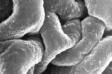
Pinholes Viewed on Silver Stain
This eyeSCANDy image is an acellular scanning EM from a biopsy with membranous glomerulonephritis, stage I showing numerous small shallow…
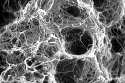
Empty Lacunar Spaces
Todays eyeSCANdy image shows acellular scanning EM showing empty lacunar spaces of a fibrocellular crescent previously occupied by cells. Photo…
Glomerulus With Fibrocellular Crescent
Its eyeSCANdy Tuesday! This image shows acellular scanning EM showing a glomerulus with a fibrocellular crescent (top). Photo credits…