Search & Filter All Posts
All Articles
(7 Results)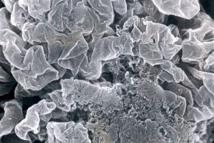
Normal Glomerular Tuft with Segmental Necrosis
This eyeSCANdy image shows acellular scanning EM with normal glomerular tuft at top and segmental necrosis at bottom. Photo courtesy…
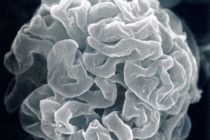
Removal of the Podocytes
Todays eyeSCANdy image shows acellular scanning EM of a normal glomerular tuft showing the subpodcocytic capillary loop basement membranes following…
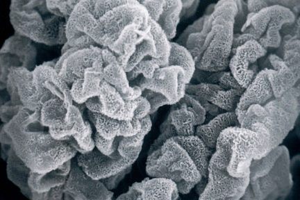
Diffuse Reticular Appearance of GBM
This eyeSCANdy image shows acellular scanning EM of a glomerulus with membranous glomerulonephritis, stage II showing diffuse reticular appearance of…
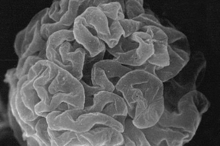
Subpodcocytic Capillary Loop Basement Membranes
This eyeSCANdy image shows acellular scanning EM of a normal glomerular tuft showing the subpodcocytic capillary loop basement membranes following…
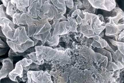
Segmental Necrosis
Todays eyeSCANdy image shows acellular scanning EM with normal glomerular tuft at top and segmental necrosis at bottom.…
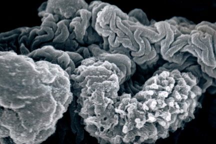
Glomerular Tuft with Collapsed GBM
This eyeSCANdy image shows portions of glomerular tuft with collapsed GBM (across top), spicular amyloid (middle right), and nodular amyloid…
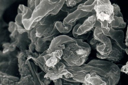
Glomerular Tuft
Today’s eyeSCANdy image shows glomerular tuft with overall intact architecture. Discrete masses of specular amyloid interrupt smooth uninvolved segments of…
