Clinical History:
Muscle biopsy from this 40-year-old patient showed morphologic and enzyme histochemical features of denervation.
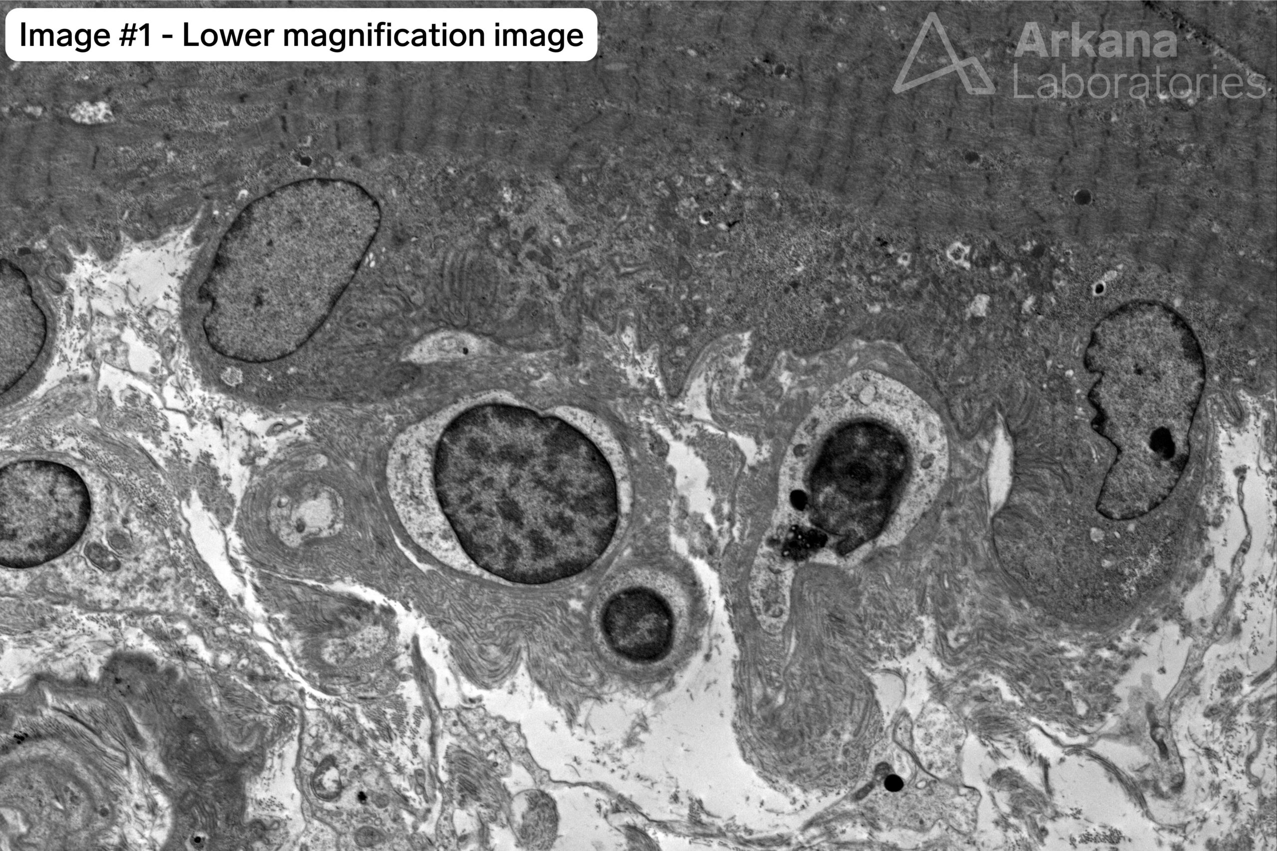
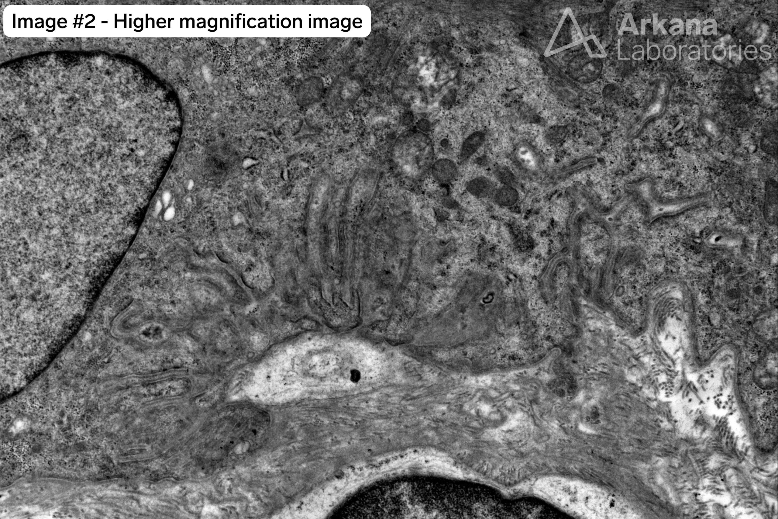
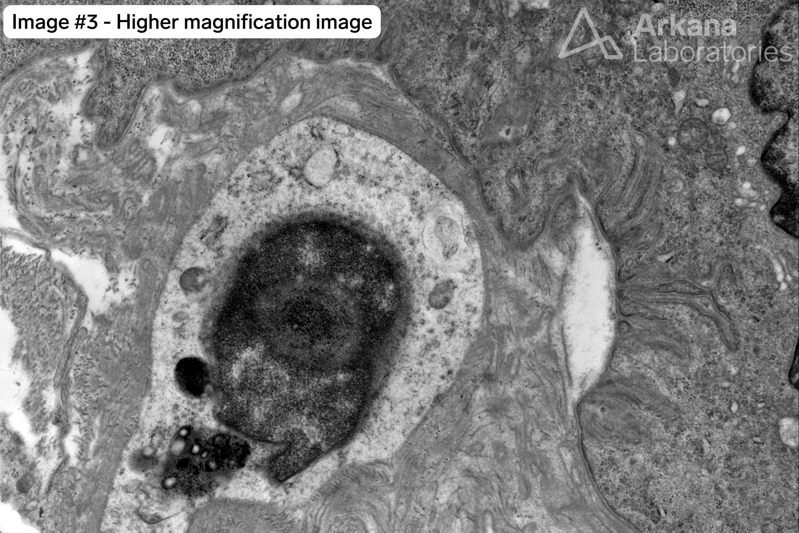
What abnormality is seen in the electron microscopic images #1 – #3 of neuromuscular junction present in this patient’s muscle biopsy?
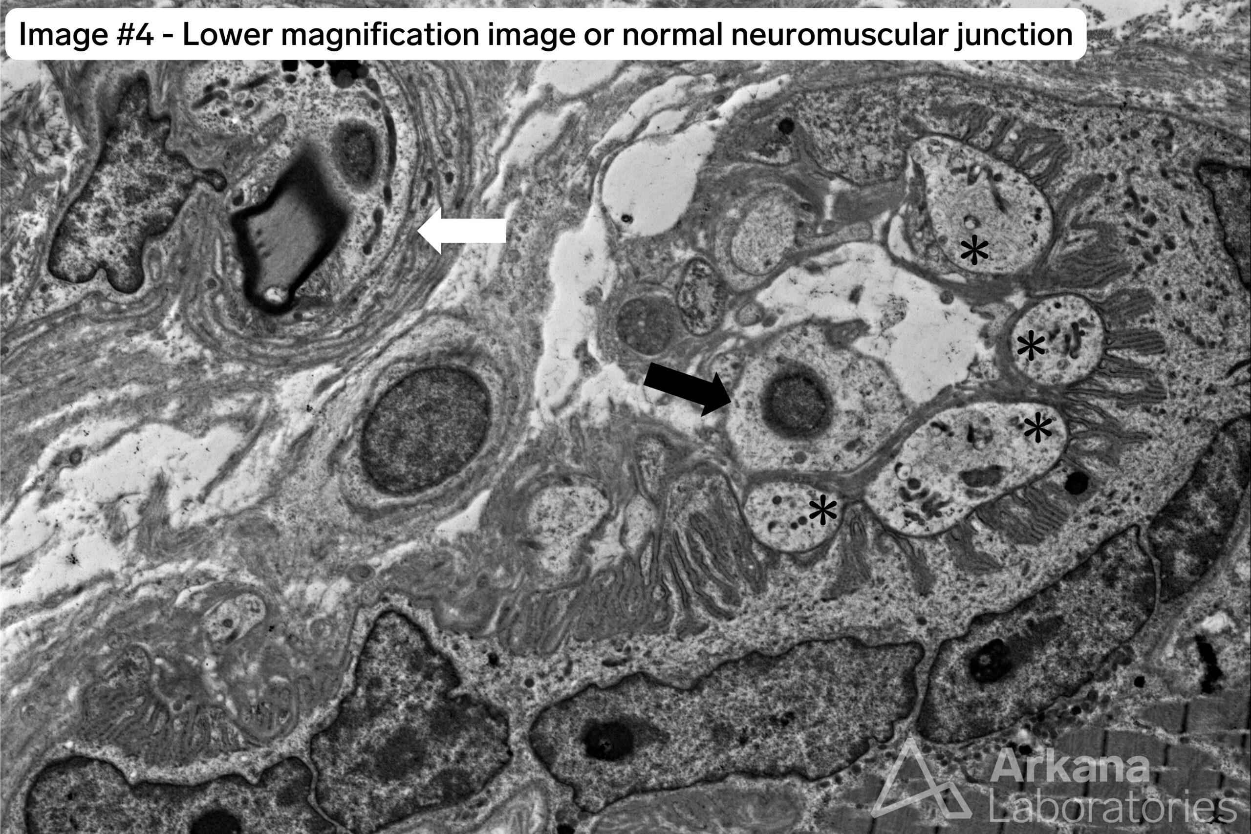
For comparison, image #4 is from a normal neuromuscular junction.
Answer:
The terminal boutons of the motor axons are missing from the neuromuscular junction (NMJ).
Image #4 of a normal NMJ shows the following:
- White arrow: small intramuscular nerve twig. The presence of these in a tissue section indicates the likely presence of NMJs in the vicinity.
- Black arrow: terminal Schwann cell.
- Asterisks symbol (*): terminal motor axon bouton. Note that these are not myelinated. Also note the adjacent motor end plate which represents a specialized area of the muscle fiber cell membrane (sarcolemma) which shows an undulating appearance due to extensive folding (junctional folds). The motor end plate is narrowly separated from the terminal axon bouton by a thin space known as the synaptic cleft.
Reference(s) / Additional Reading:
Quick note: This post is to be used for informational purposes only and does not constitute medical or health advice. Each person should consult their own doctor with respect to matters referenced. Arkana Laboratories assumes no liability for actions taken in reliance upon the information contained herein.