What is your diagnosis based on the EM image provided?
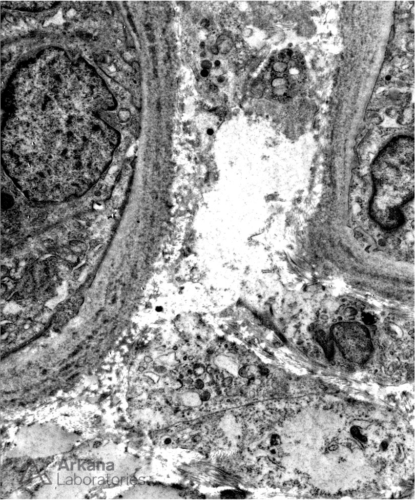
The electron microscopy image provided is that of portions of proximal tubules and demonstrates “powdery” or “pellet-like” deposits along the tubular basement membrane consistent with a diagnosis of light chain deposition disease (LCDD), the most common subtype of monoclonal immunoglobulin deposition disease (MIDD). By immunofluorescence, the case revealed diffuse, linear, 3+ staining for lambda light chain along tubular basement membranes as well as glomerular basement membranes while kappa was completely negative.
As the name implies, MIDD is caused by the systemic deposition of monoclonal immunoglobulin along tubular and glomerular basement membranes and within the glomerular mesangium. The monoclonal immunoglobulin is typically of the light chain type but can also be due to deposition of an abnormal heavy chain (HCDD) or even both, light and heavy chains (LHCDD). Of note, while most patients will demonstrate an abnormal SPEP/UPEP at the time of diagnosis, approximately 15-20% will have an undetectable paraprotein at that time. However, all patients should demonstrate an abnormal serum free light chain assay at the time of diagnosis.
Quick note: This post is to be used for informational purposes only and does not constitute medical or health advice. Each person should consult their own doctor with respect to matters referenced. Arkana Laboratories assumes no liability for actions taken in reliance upon the information contained herein.