These are the C1q and C3 stains of the biopsy of a 13-year-old female with hematuria and proteinuria. Given these findings, what is your main differential diagnosis.
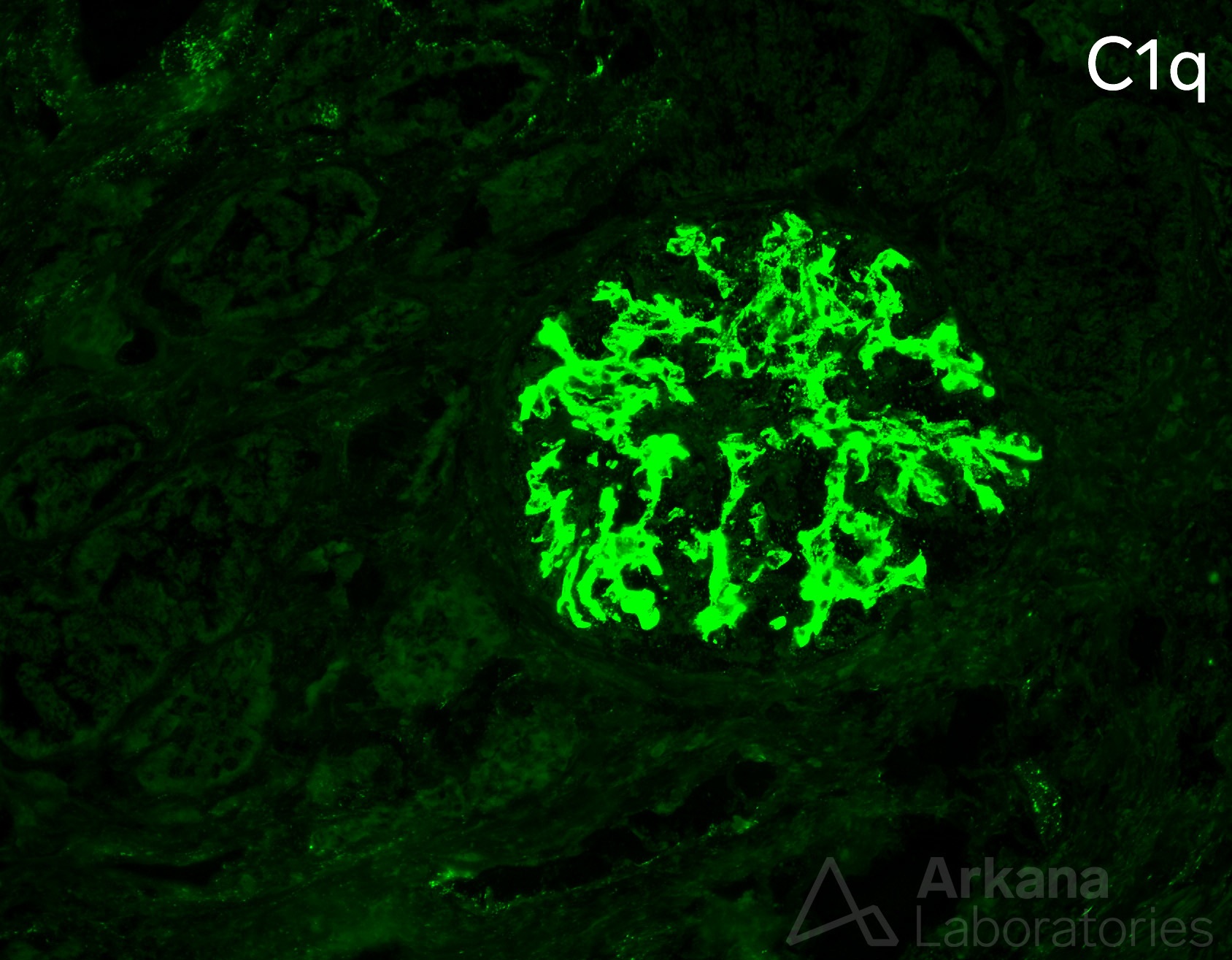
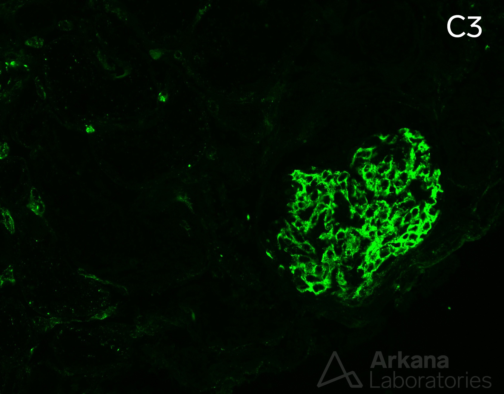
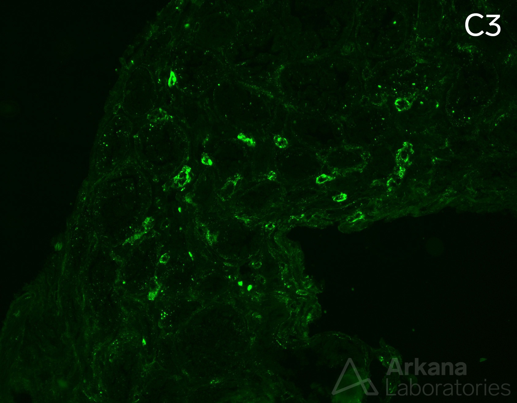
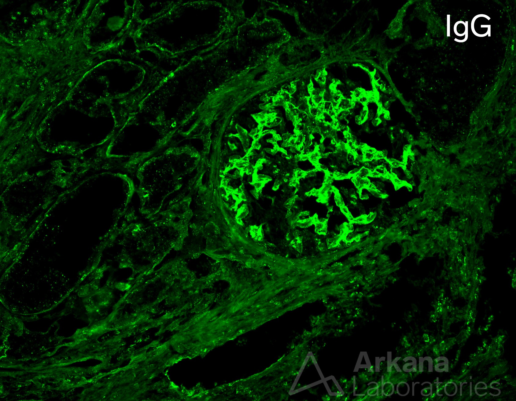
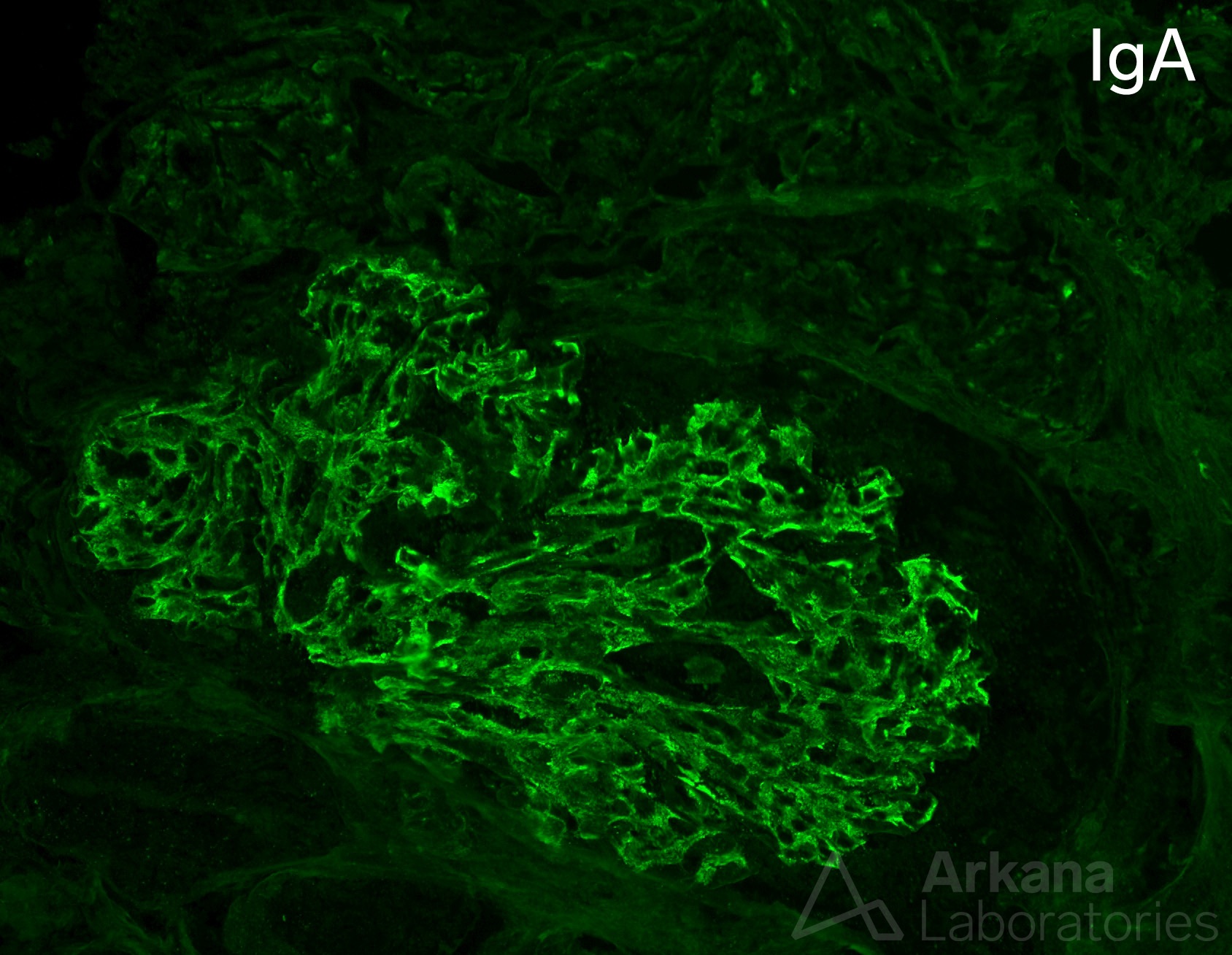
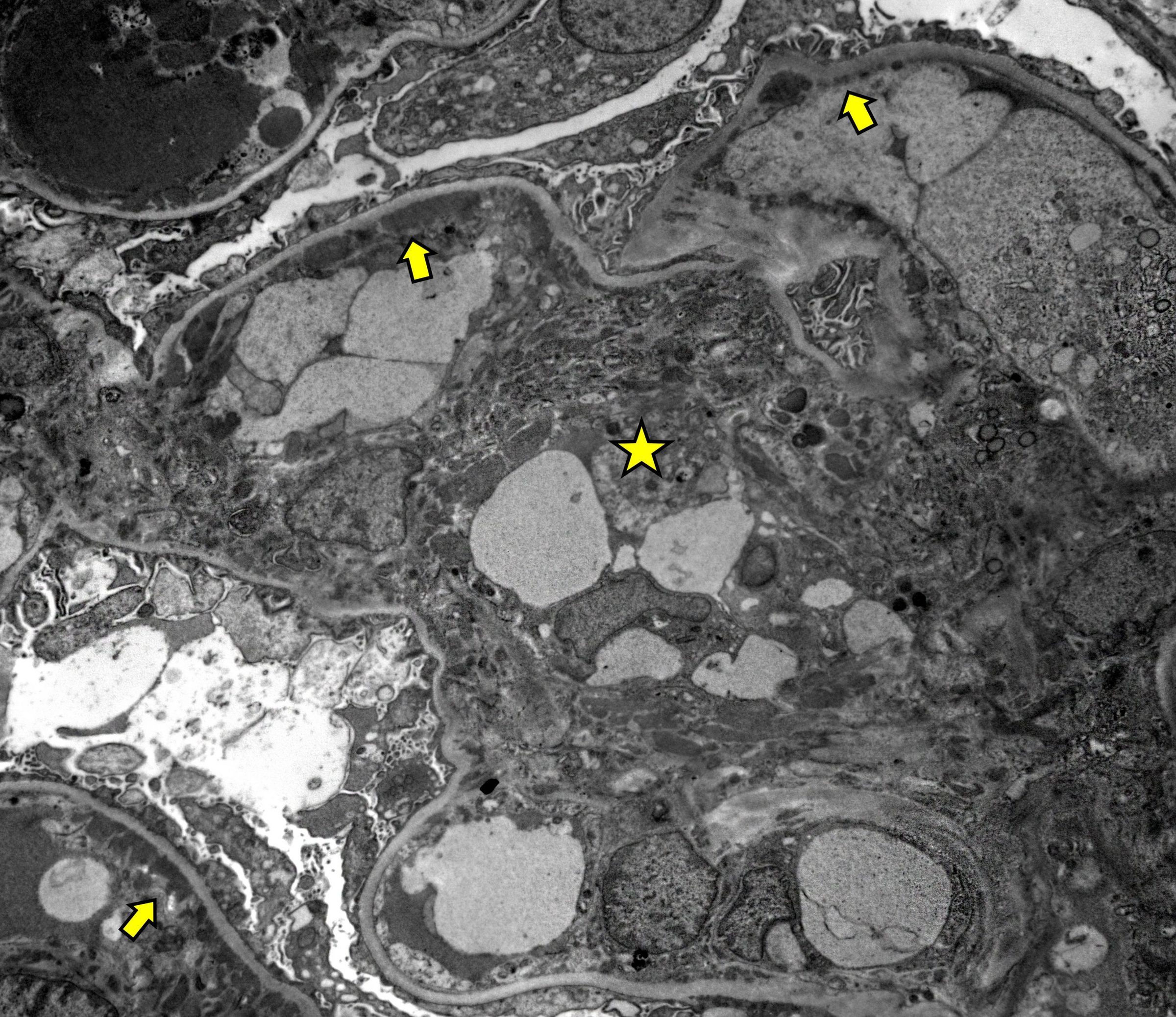
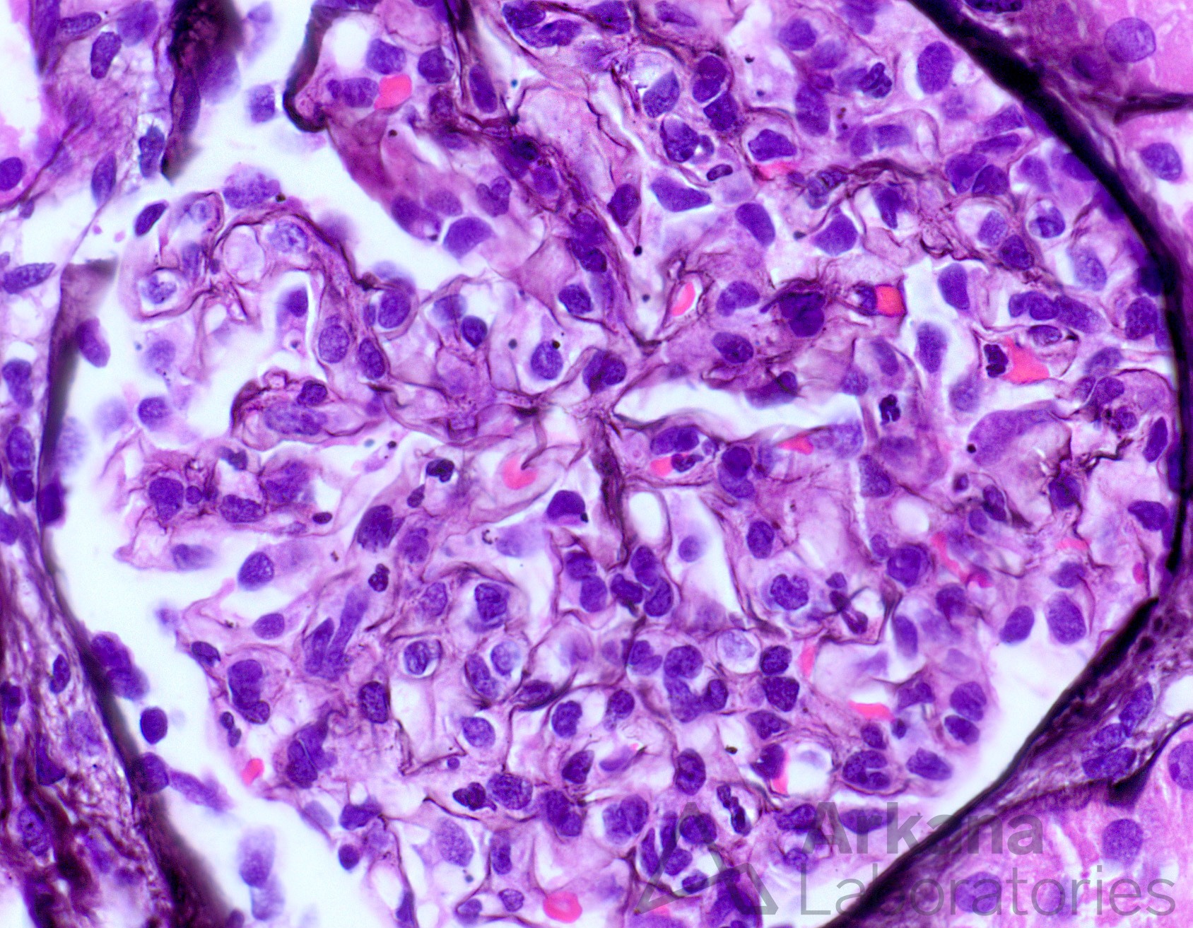
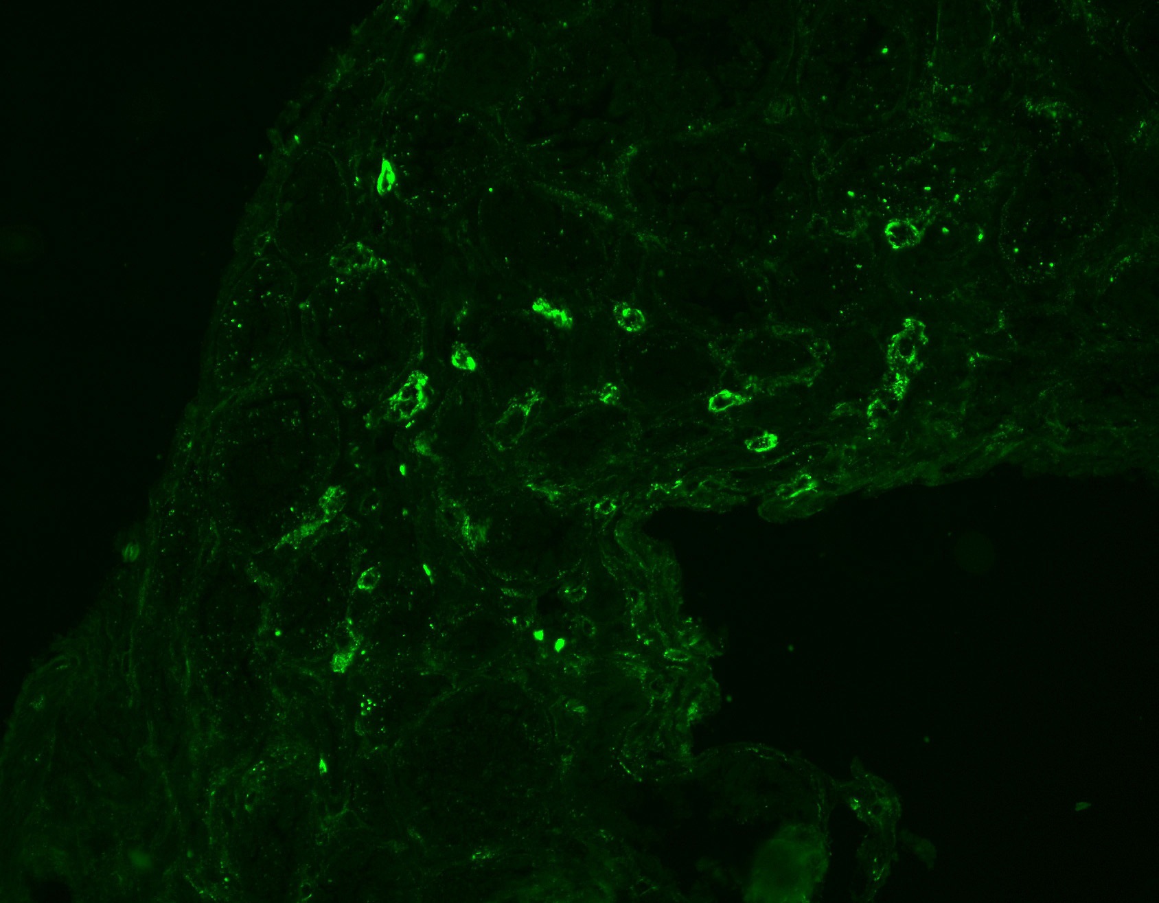
The biopsy shows mesangial and capillary wall staining for C3 and C1q, as well as peritubular capillary staining for C3. The presence of strong C3 and C1q (both 3+) excludes the possibility of C3 Glomerulopathy, and the presence of extra-glomerular staining (peritubular capillary) suggests underlying autoimmune diseases, such as SLE. There was also IgG and IgA staining. By light microscopy, there was endocapillary hypercellularity, and no GBM lucencies or spikes. Ultrastructurally, numerous mesangial (star) and subendothelial (arrows) electron dense deposits were present. Indeed, the patient had SLE and this was diagnosed as a class IV lupus nephritis.
Quick note: This post is to be used for informational purposes only and does not constitute medical or health advice. Each person should consult their own doctor with respect to matters referenced. Arkana Laboratories assumes no liability for actions taken in reliance upon the information contained herein.