In a patient with progressively rising serum creatinine and a history of CABG 2 months prior, what is your diagnosis?
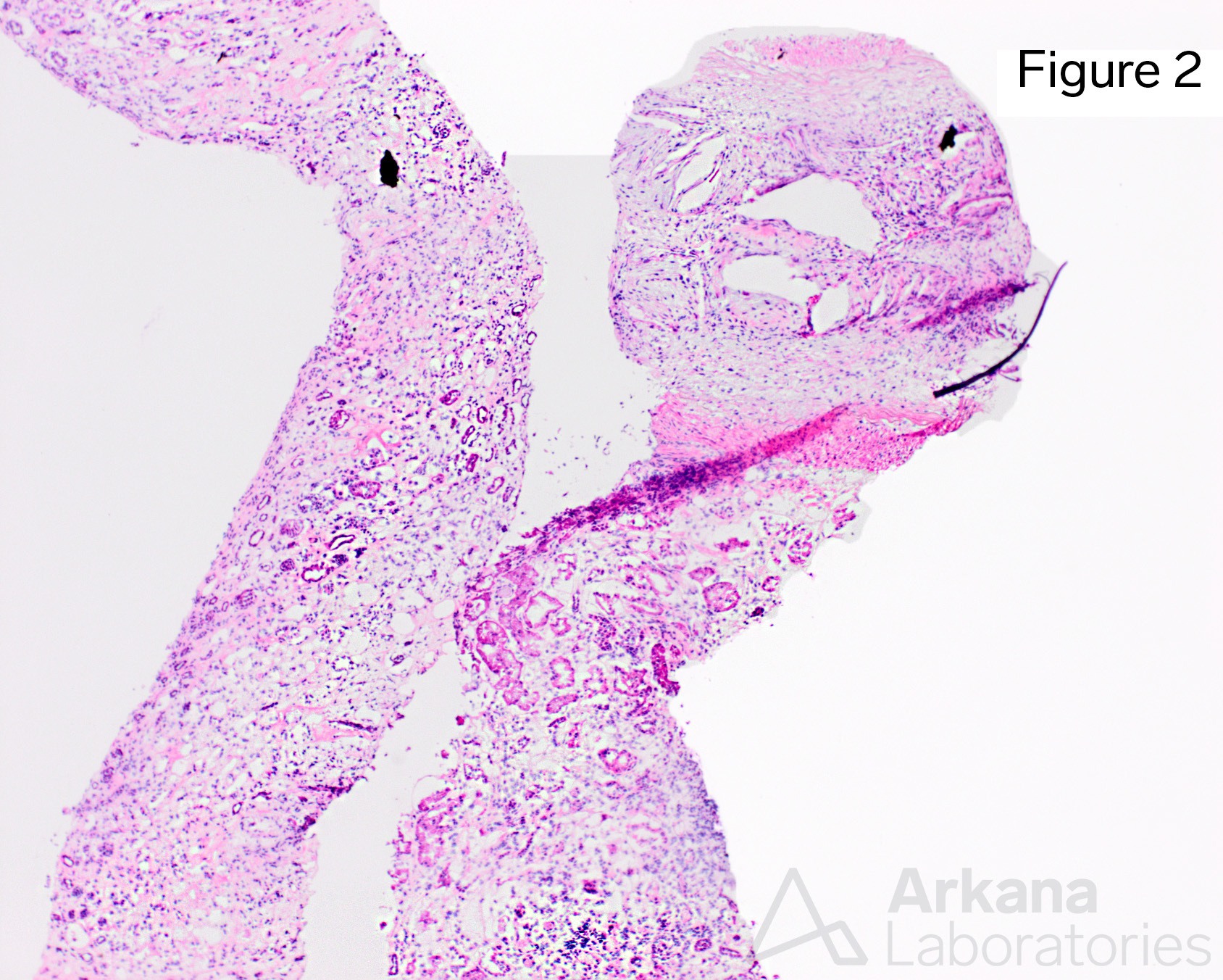
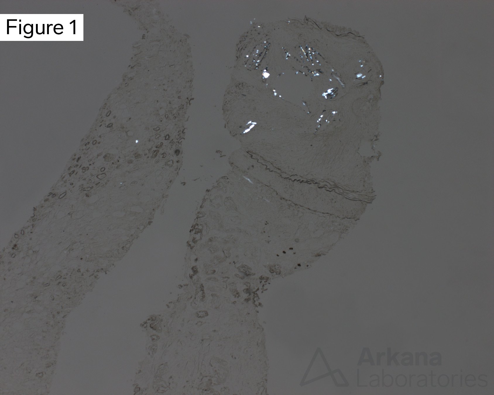
The images are from frozen sections of the cores submitted for immunofluorescence. By H&E staining, a large caliber artery can be seen, with numerous intraluminal clefts, surrounded by fibroblasts and inflammatory cells. In an albumin-stained section viewed under polarization on bright field, birefringent crystals are seen in the areas corresponding to the clefts, consistent with cholesterol emboli. Cholesterol emboli do not dissolve in the immunofluorescence sections as those do not undergo formalin fixation and paraffin embedding; therefore, not only clefts but the actual crystals can be seen if those sections are examined under polarized light.
In this case, no arteries in the light microscopy sections had cholesterol emboli; highlighting the importance of examining also the immunofluorescence frozen sections when cholesterol emboli are suspected clinically.
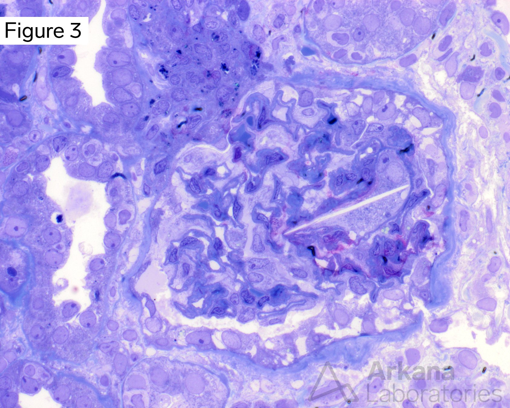
On toluidine blue sections of the material submitted for ultrastructural examination, a single needle-shaped cleft was seen, presumably within a macrophage lodged in a glomerular capillary (Figure 3).
Quick note: This post is to be used for informational purposes only and does not constitute medical or health advice. Each person should consult their own doctor with respect to matters referenced. Arkana Laboratories assumes no liability for actions taken in reliance upon the information contained herein.