What is your diagnosis?
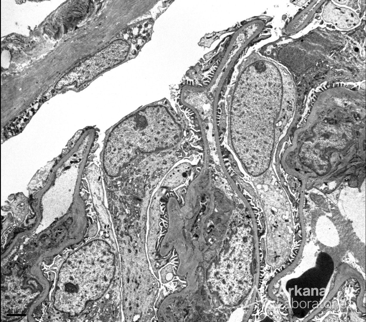
The EM photomicrograph shows capillary walls that are uniform and of normal thickness with no electron dense immune complex-type deposits noted within the capillary walls or mesangium. Additionally, numerous podocyte cell bodies are present and unremarkable and epithelial foot processes show no significant effacement. At the top left of the image you can even see Bowman’s capsule and unremarkable parietal epithelium. These findings are those of a normal glomerulus by electron microscopy. No pathologic changes are present in this image.
Quick note: This post is to be used for informational purposes only and does not constitute medical or health advice. Each person should consult their own doctor with respect to matters referenced. Arkana Laboratories assumes no liability for actions taken in reliance upon the information contained herein.