Search & Filter All Posts
All Articles
(9 Results)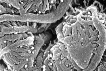
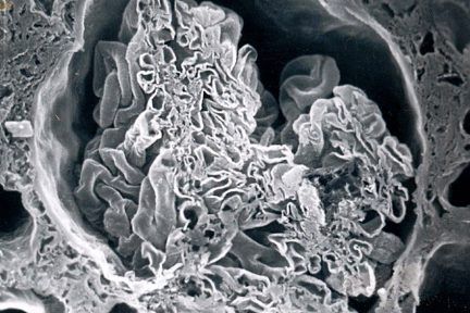
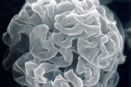
Normal Glomerulus
This scanning EM of a normal glomerulus shows a glomerular capillary loop with interdigitating podocyte foot processes. Photo credits to…
Interdigitating Podocyte Foot Processes
This eyeSCANdy scanning EM image of a normal glomerulus shows a glomerular capillary loop with interdigitating podocyte foot processes. Photo…
Origin of the Proximal Tubule From Bowman’s Capsule
This eyeSCANdy acellular scanning EM image shows a glomerulus in cross-section through the macular densa and shows the origin of…
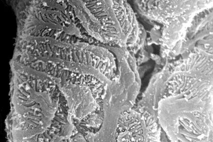
Macula Densa Basement Membrane Tunnels
Todays eyeSCANdy image shows a normal glomerulus sectioned through the vascular pole showing the macula densa basement membrane tunnels. Photo…
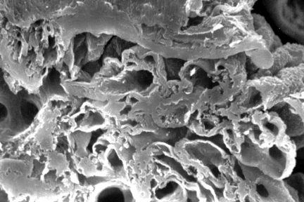
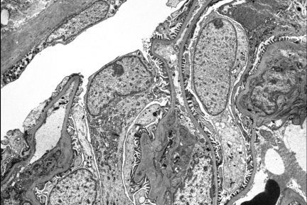
Removal of the Podocytes
Todays eyeSCANdy image shows acellular scanning EM of a normal glomerular tuft showing the subpodcocytic capillary loop basement membranes following…
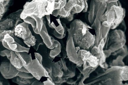
Glomerular Tuft with Intact Architecture
Today’s eyeSCANdy image shows glomerular tuft with overall intact architecture. Discrete masses of specular amyloid (long arrows) interrupt smooth uninvolved…

Malpighi Glomerulus
How long have we known about glomeruli? Todays teaching point is offered in honor of Marcello Malpighi (1628-1694), the Italian…
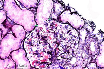
Normal Histology
This is a normal glomerulus by light microscopy using the Jones silver stain. This section is fortuitously cut through the…