

Clinical History:
This 80-year-old patient presented with complaints of left lower leg muscle pain clinically suspicious for myositis. Their past medical history was significant severe venous stasis involving both lower legs with focal nonhealing ulcers. Laboratory studies showed positive ANA and CRP. Intraoperatively, muscle tissue had an abnormal brownish discoloration and did contract when stimulated.
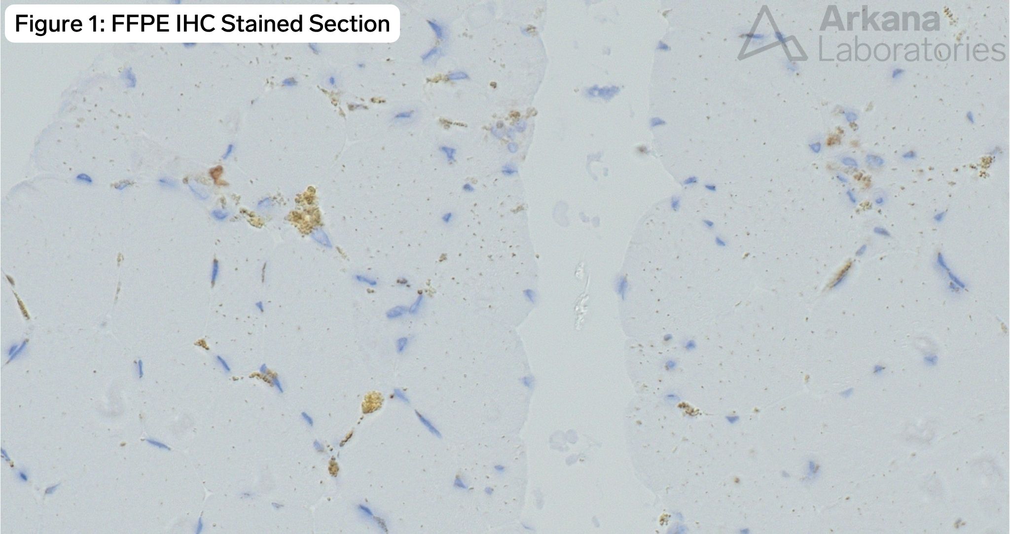
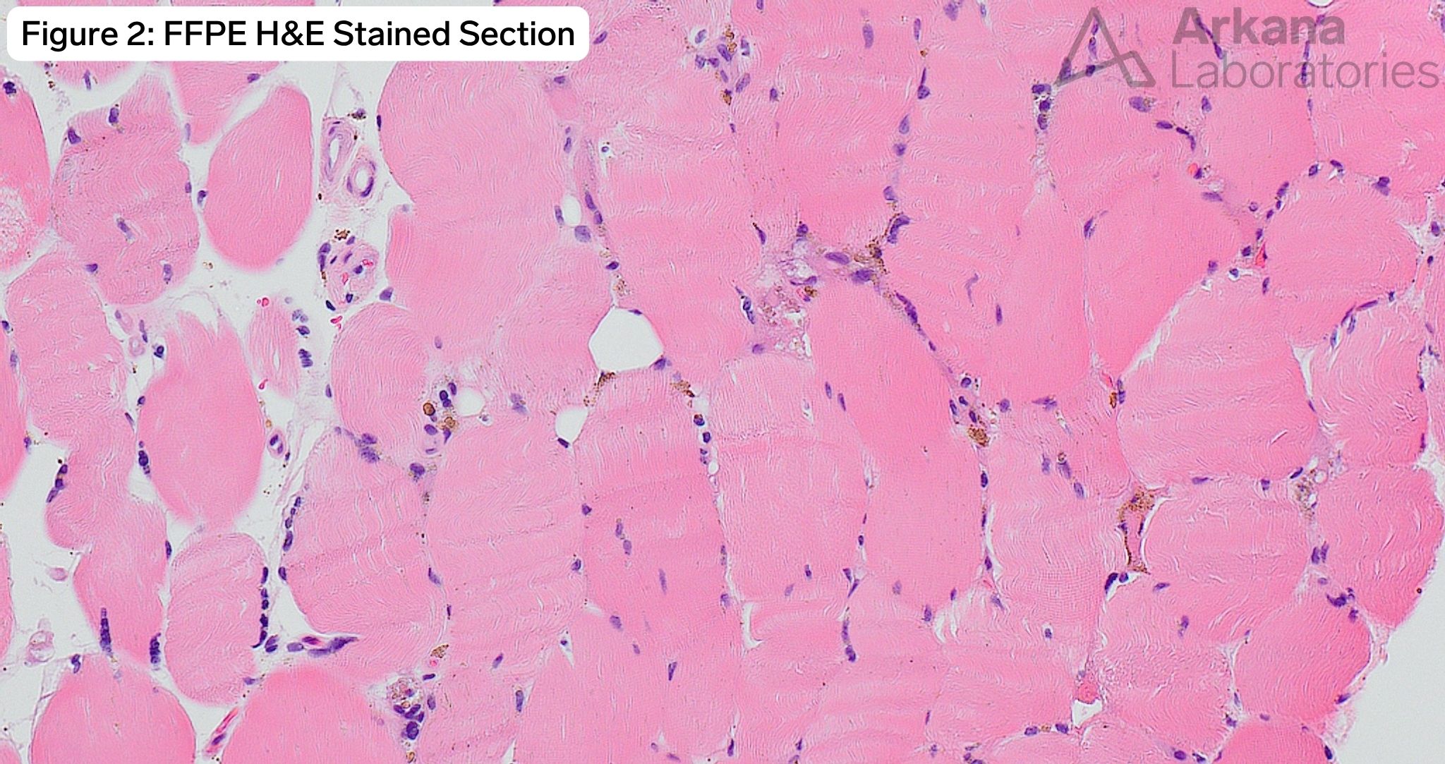
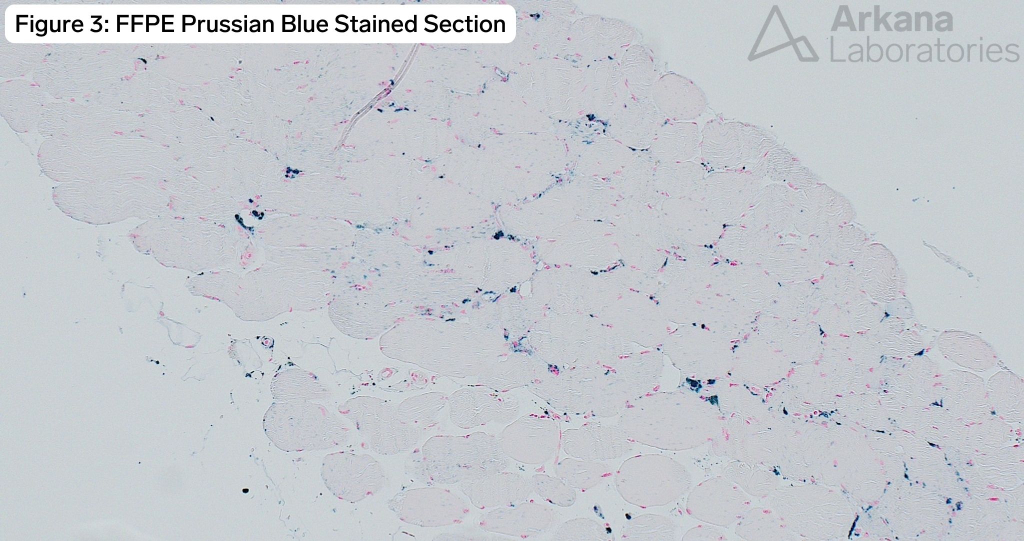
What is the brownish material seen on the immunohistochemically stained section? An H&E and Prussian Blue stain image are also supplied.
A. Positive immunostaining
B. Hemosiderin
C. Calcium
D. Endogenous peroxidase
Answer:
Hemosiderin pigment
- There is marked granular to globular pericapillary hemosiderin deposition. This is well-demonstrated on the H&E and Prussian Blue (Iron) stained sections.
- Hemosiderin is one the of pigmented materials that may render the interpretation of immunohistochemical stains using DAB (3,3′-diaminobenzidine tetrahydrochloride; brown chromagen) difficult. This can be resolved with the use of a red chromagen.
- In this case no positive immunohistochemical staining was present. The granular material represents hemosiderin.
- Dystrophic calcium deposition characteristically has a somewhat blue-black tinctoral quality.
- Inadequate quenching of endogenous peroxidase may result in non-specific background immunostaining of some tissue/cell types (for example red blood cells) when using HRP-conjugate antibodies.
Quick note: This post is to be used for informational purposes only and does not constitute medical or health advice. Each person should consult their own doctor with respect to matters referenced. Arkana Laboratories assumes no liability for actions taken in reliance upon the information contained herein.


