


Clinical History:
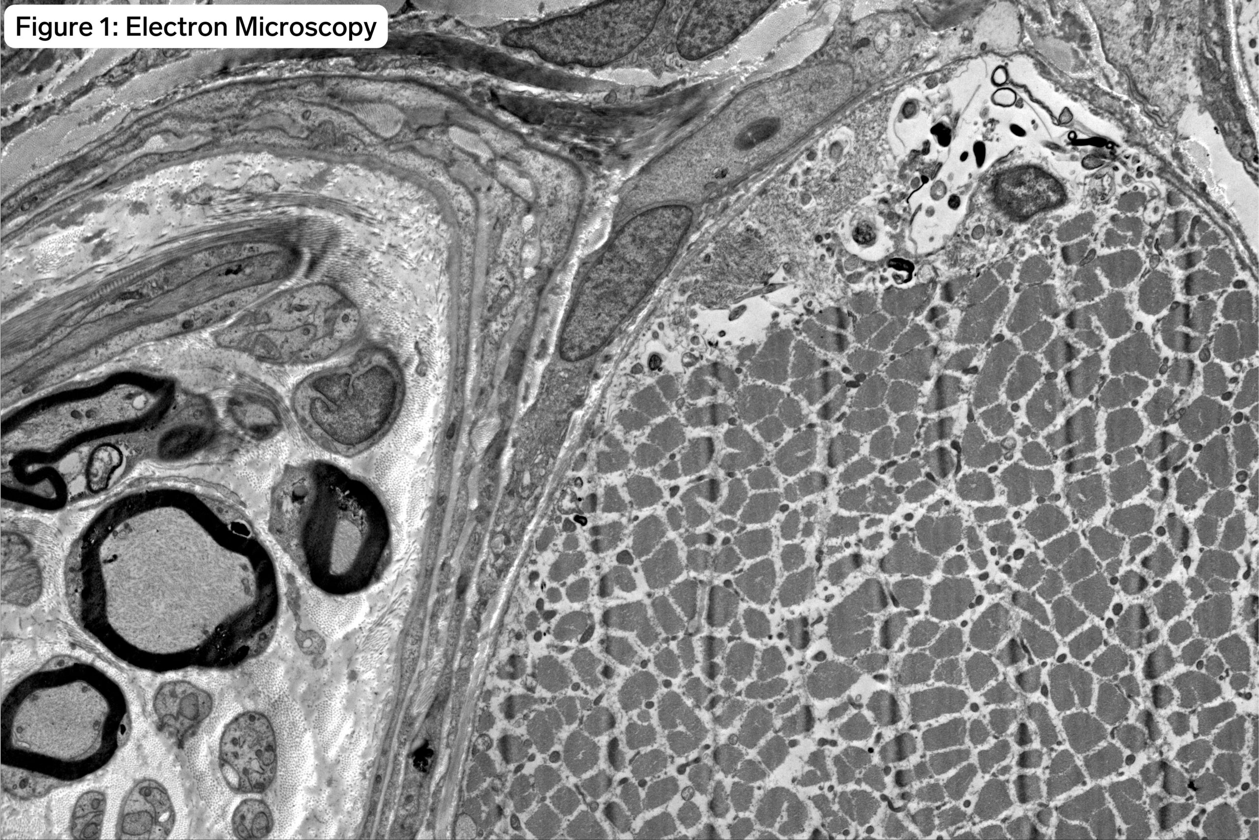
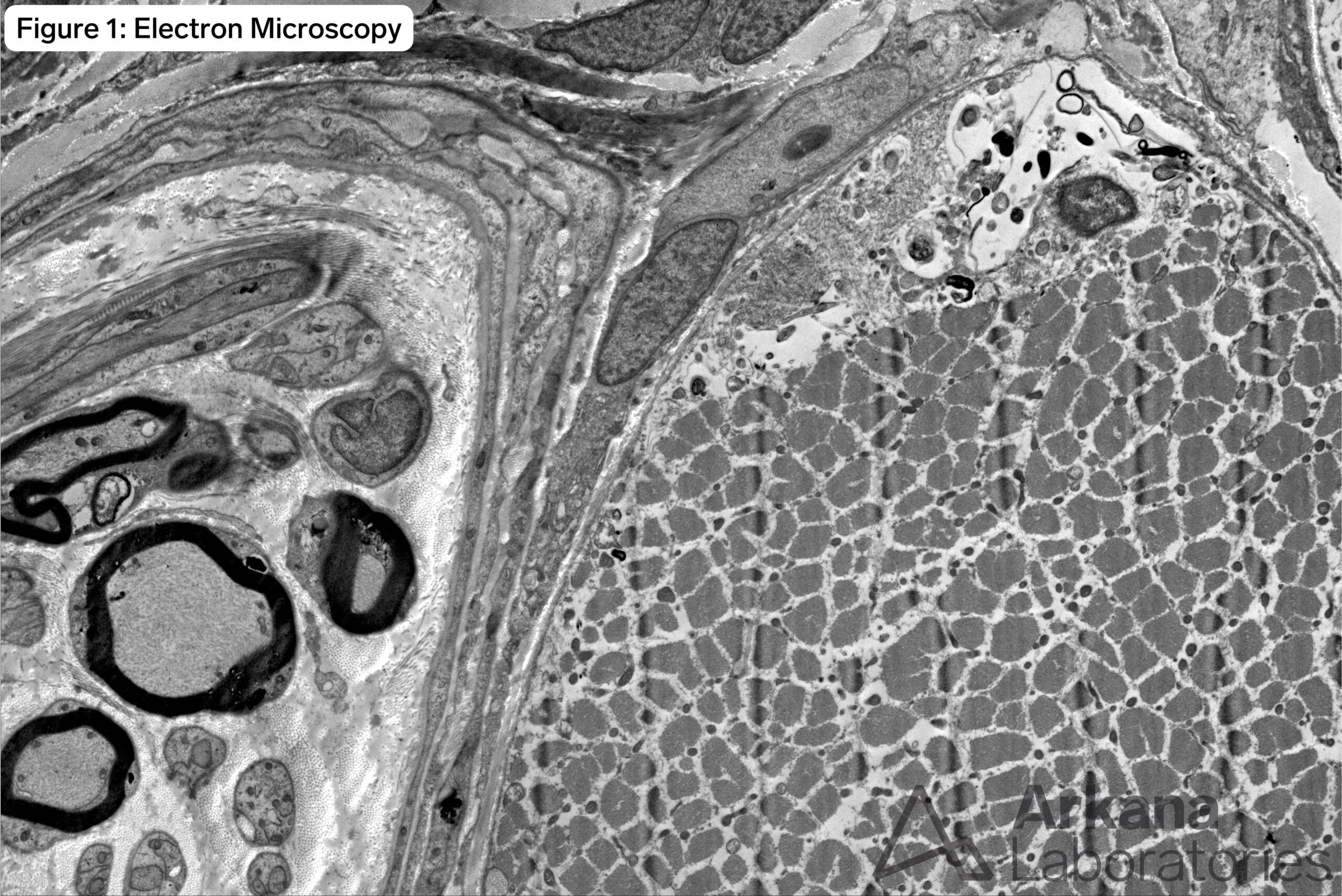
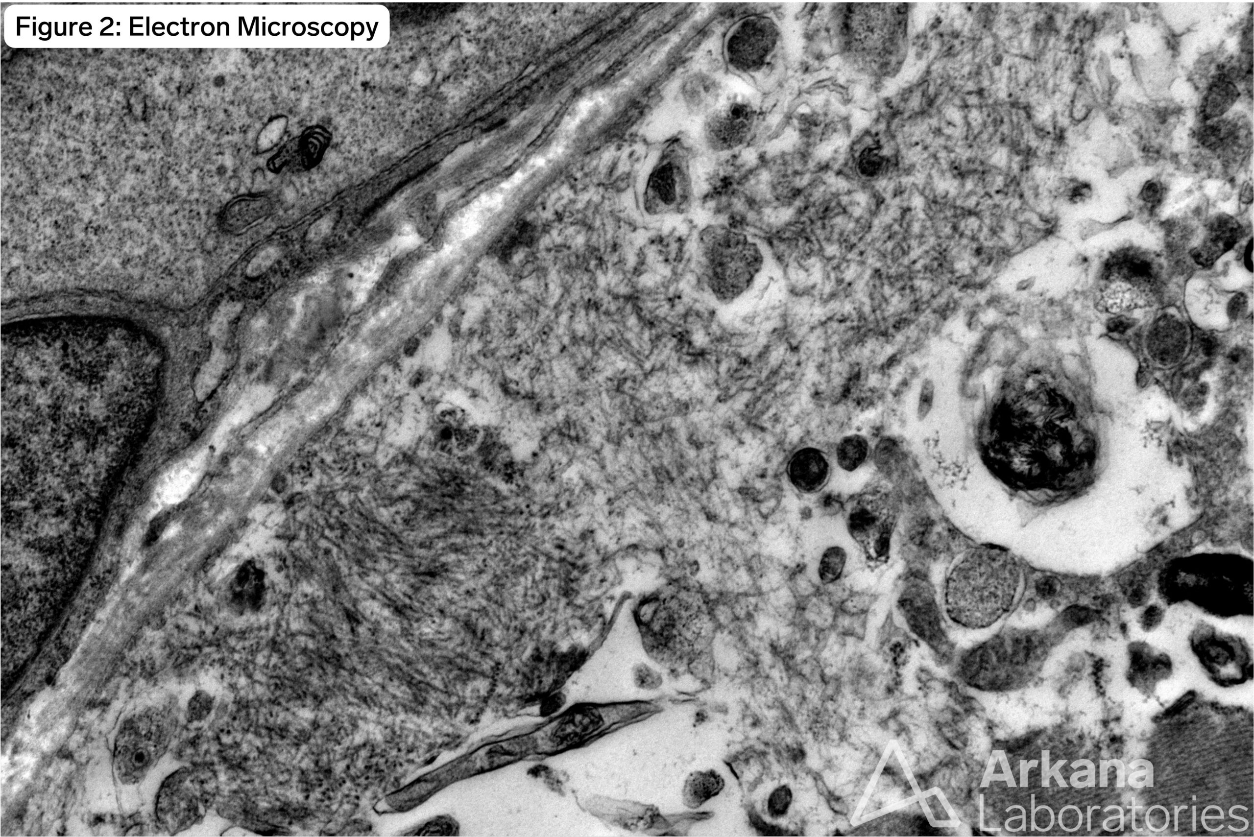
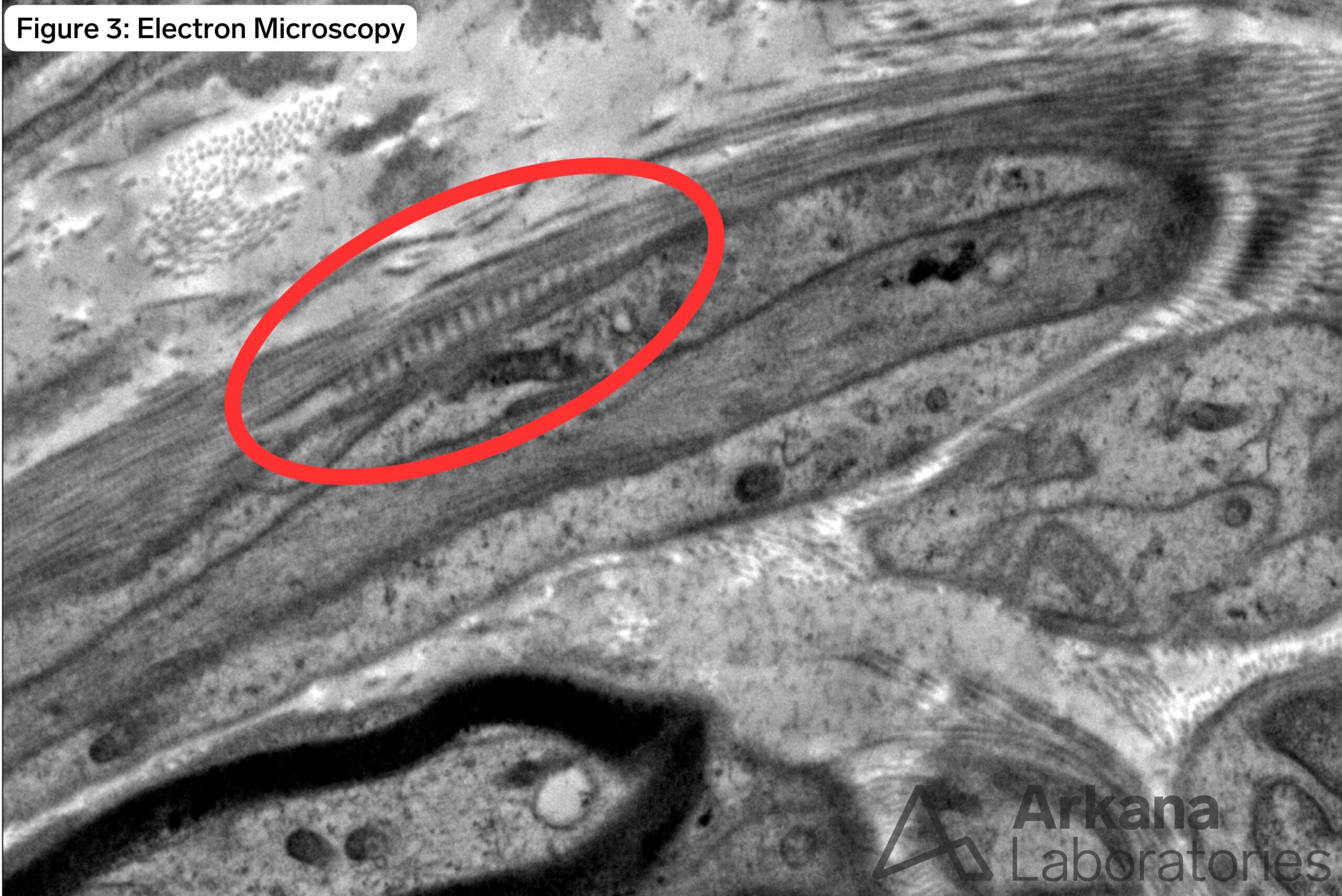
No clinical history was available for this 70-year-old patient. Muscle biopsy showed features of a chronic active inflammatory myopathy with rimmed-vacuoles, and associated congophilic-TDP43-p62 positive intrasarcoplasmic material. Electron microscopy confirmed the presence of amyloid-like fibrillary inclusions (see images #1 and #2 below) supporting a pathologic diagnosis of sporadic Inclusion Body Myositis (sIBM). An incidental finding was noted within the endoneurium of a small peripheral nerve present in the muscle biopsy tissue available for electron microscopy (see image #3).
Which of the following is this incidental finding called?
A. Luse body
B. Long spaced collagen
C. “Raccoon tail”
D. All of the above
Answer:
D. All of the above
Luse bodies (aka long space collagen or “raccoon tails”) are an incidental nonspecific finding which may be seen in peripheral nerve biopsy and benign peripheral nerve sheath tumors, but also in a variety of other tissues (both normal and pathologic). The fusiform collection of collagen has a characteristic banding pattern (periodicity) that is greater than normal collagen fibrils.
Reference(s) / Additional Reading:
- Paige MF, Rainey JK, Goh MC. Fibrous long spacing collagen ultrastructure elucidated by atomic force microscopy. Biophys J. 1998 Jun;74(6):3211-6. doi: 10.1016/S0006-3495(98)78027-0. PMID: 9635774; PMCID: PMC1299661.
Quick note: This post is to be used for informational purposes only and does not constitute medical or health advice. Each person should consult their own doctor with respect to matters referenced. Arkana Laboratories assumes no liability for actions taken in reliance upon the information contained herein.


