This 55 year-old patient presented with generalized muscle weakness and normal CPK.
Which of the following does the electron microscopic image in Figure #1 show?
A. Nemaline rod
B. Satellite cell
C. Mitotic figure
D. Pseudoinclusion

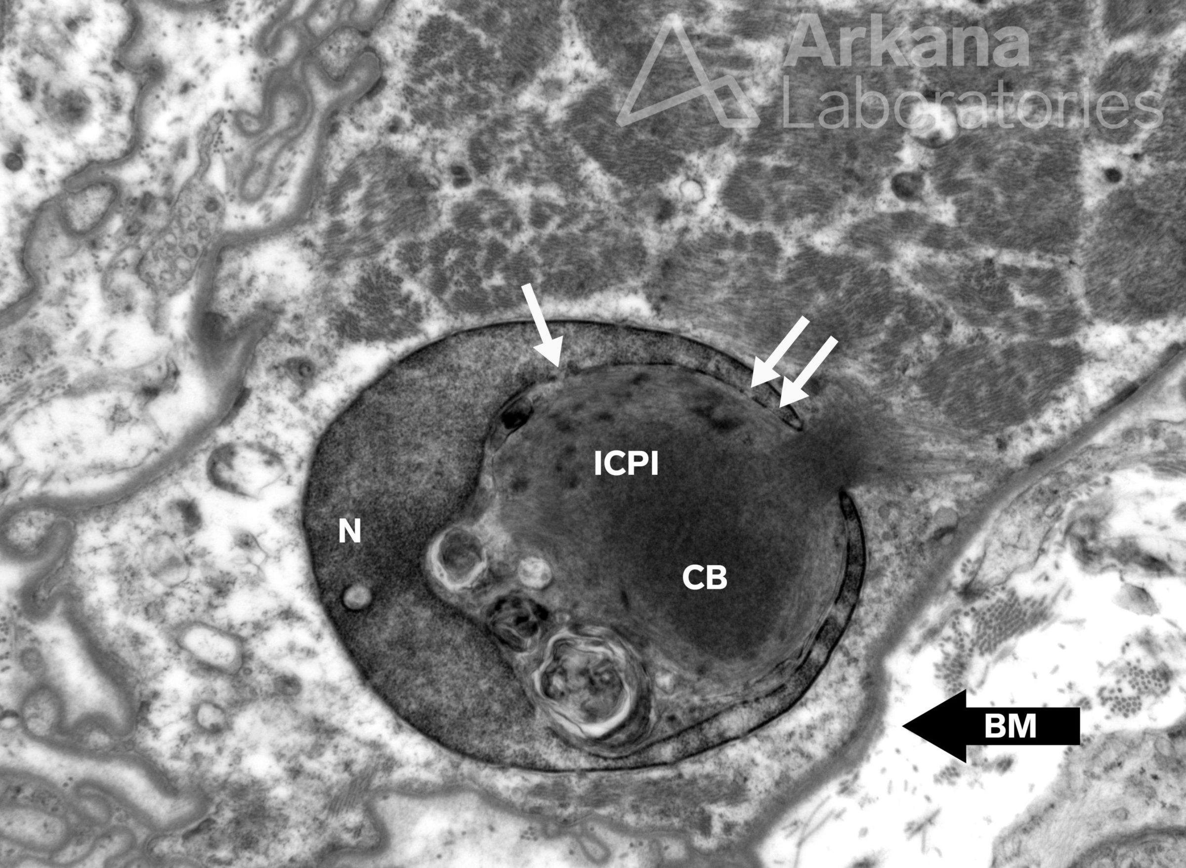
Figure 1: Electron microscopy 12000x original magnification
Answer: Pseudoinclusion
The image shows an intranuclear cytoplasmic pseudoinclusion. This represents sectioning through a nodular protrusion of cytoplasm (sarcoplasm in this case) that indents the nucleus….. visualize pushing your fist into an inflated balloon and then taking a cross section.
These are called “pseudoinclusions” because they are not actually inside the nucleus. This fact is made visible by the intact heterochromatin clinging to the nuclear membrane around the entire “C” shaped contour of the nucleus. If you look closely, other visible features include the double membrane of the nucleus and the nuclear pores.
This particular image is unusual in that it shows a cytoplasmic body occupying the intranuclear cytoplasmic pseudoinclusion. Note how its filaments focally extend past the contour of the nucleus.
This is an unusual morphologic finding of no known clinical significance. For some reason, findings like these seem cling to my memory the best.
Quick note: This post is to be used for informational purposes only and does not constitute medical or health advice. Each person should consult their own doctor with respect to matters referenced. Arkana Laboratories assumes no liability for actions taken in reliance upon the information contained herein.