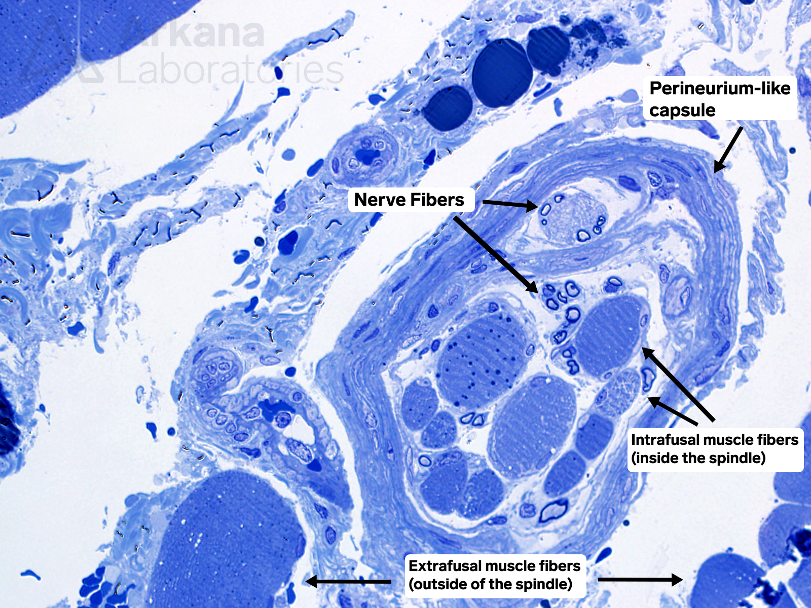
Toluidine blue-stained thick section prepared from glutaraldehyde-fixed EPON embedded tissue (400x original magnification)
Muscle biopsy was performed on this 5-year old male to evaluate potential causes for mild unilateral muscle weakness. What is the structure in the right half of the image?
A. Intramuscular parasite
B. Nerve twig
C. Ring fiber
D. Muscle fiber spindle
Answer: Muscle fiber spindle
The muscle fiber spindle is a mechanoreceptor (stretch receptor) for muscle, and helps to coordinate agonist and antagonist muscle contraction/relaxation. The figure also shows perineurium-like capsule, nerve fibers, intrafusal muscle fibers (inside the spindle), and extrafusal muscle fibers (outside of the spindle).
References
Banks RW. The innervation of the muscle spindle: a personal history. J Anat. 2015 Aug;227(2):115-35. doi: 10.1111/joa.12297. Epub 2015 Jun 19. PMID: 26095428; PMCID: PMC4523316.
Kröger S, Watkins B. Muscle spindle function in healthy and diseased muscle. Skelet Muscle. 2021 Jan 7;11(1):3. doi: 10.1186/s13395-020-00258-x. PMID: 33407830; PMCID: PMC7788844.
Thornell LE, Carlsson L, Eriksson PO, Liu JX, Österlund C, Stål P, Pedrosa-Domellöf F. Fibre typing of intrafusal fibres. J Anat. 2015 Aug;227(2):136-56. doi: 10.1111/joa.12338. PMID: 26179023; PMCID: PMC4523317.
Walkowski AD, Munakomi S. Monosynaptic Reflex. 2021 Feb 8. In: StatPearls [Internet]. Treasure Island (FL): StatPearls Publishing; 2021 Jan–. PMID: 31082072.
Quick note: This post is to be used for informational purposes only and does not constitute medical or health advice. Each person should consult their own doctor with respect to matters referenced. Arkana Laboratories assumes no liability for actions taken in reliance upon the information contained herein.