Considering the clinical vignette from last week (elderly male with proximal muscle weakness). Staining the skeletal muscle biopsy with myoadenylate deaminase (MAD) can give the neuropathologist more information than just it’s presence or absence, as may be seen in adenosine monophosphate deaminase (AMPD) deficiency. Using the images, what other structures or ultrastructural clues can be yielded from myoadenylate deaminase (MAD) staining in this skeletal muscle biopsy?
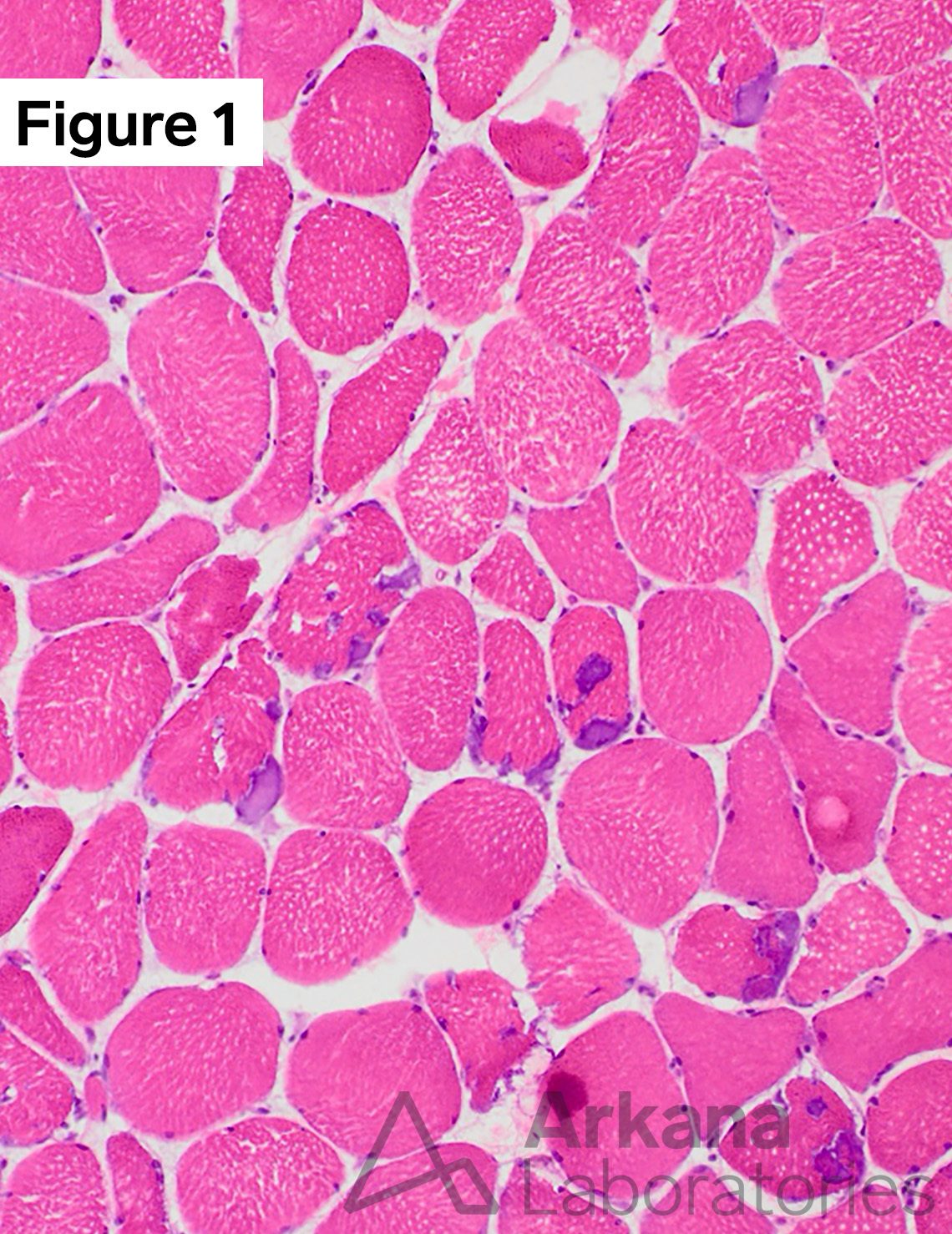
Frozen sections of skeletal muscle with Tubular Aggregate Myopathy (TAM) demonstrating a cluster of muscle fibers with ovoid eccentric to subsarcolemmal basophilic (blue) inclusions (1) with routine hematoxylin and eosin (H&E) stain, and another cluster of muscle fibers on a deeper section show inclusion material with dense dark navy blue staining character (2) against the light blue background of an intact myoadenylate deaminase (MAD) preparation. (H&E and MAD stains: original magnification 100x, each cropped for size).
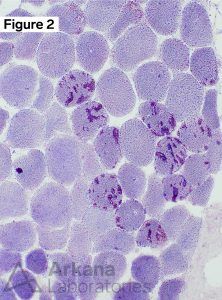
Answer: Myoadenylate deaminase (MAD) preparation can be helpful to the neuropathologist in other ways besides its presence or absence of staining. The utility of MAD staining includes clues to the underlying ultrastructural abnormality in Tubular Aggregate Myopathy (TAM), metabolic myopathy (mitochondria), and inflammatory myopathy (rimmed vacuoles of inclusion body myositis). MAD staining is not helpful in identifying metabolic myopathies with abnormal glycogen or lipid accumulations.
Quick note: This post is to be used for informational purposes only and does not constitute medical or health advice. Each person should consult their own doctor with respect to matters referenced. Arkana Laboratories assumes no liability for actions taken in reliance upon the information contained herein.