Clinical History:
Muscle biopsy was performed on this 60-year-old patient to evaluate for subjective complaints of muscle weakness. Several small intramuscular nerve twigs were noted on glutaraldehyde-fixed toluidine blue stained thick sections.
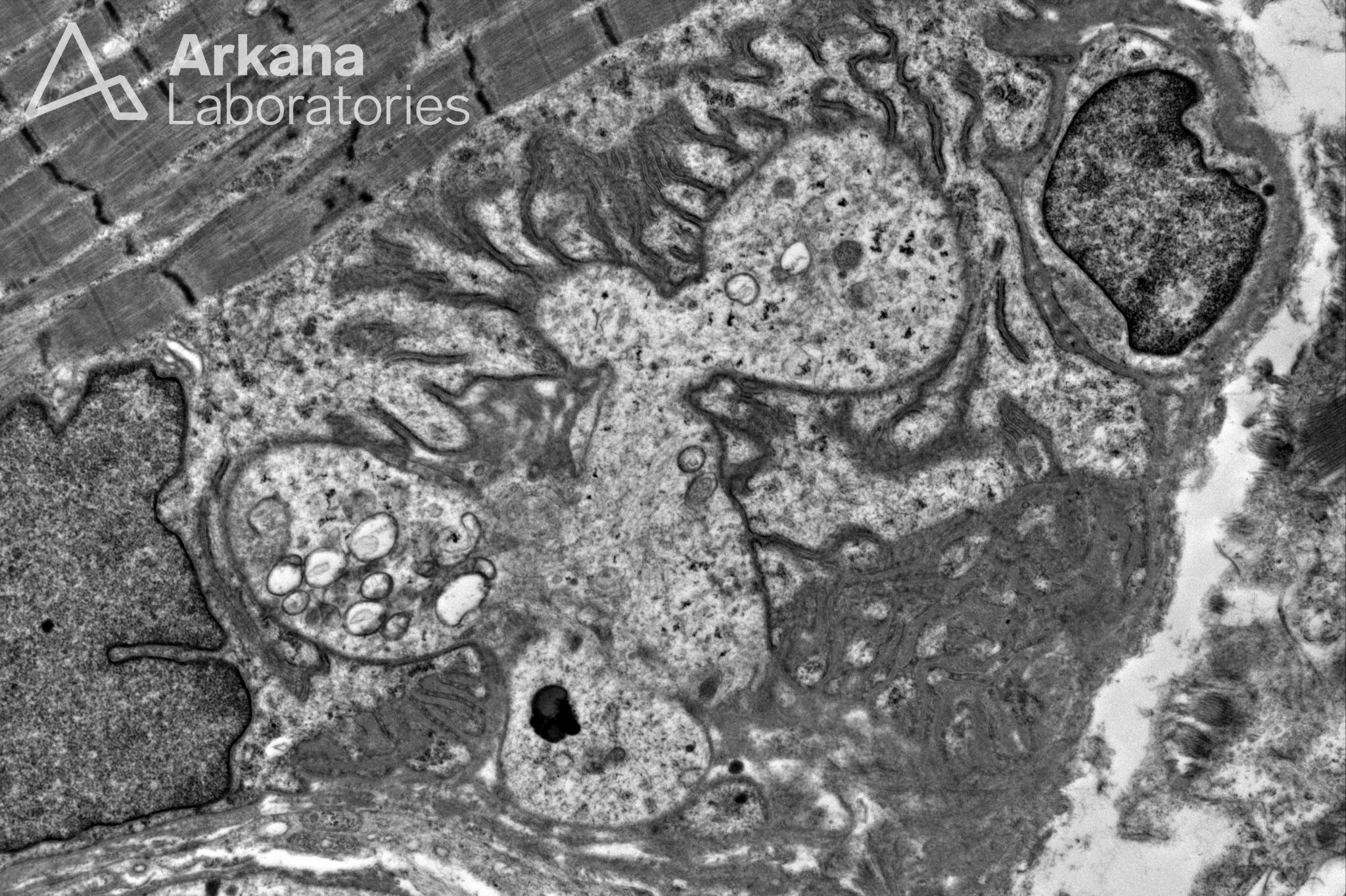
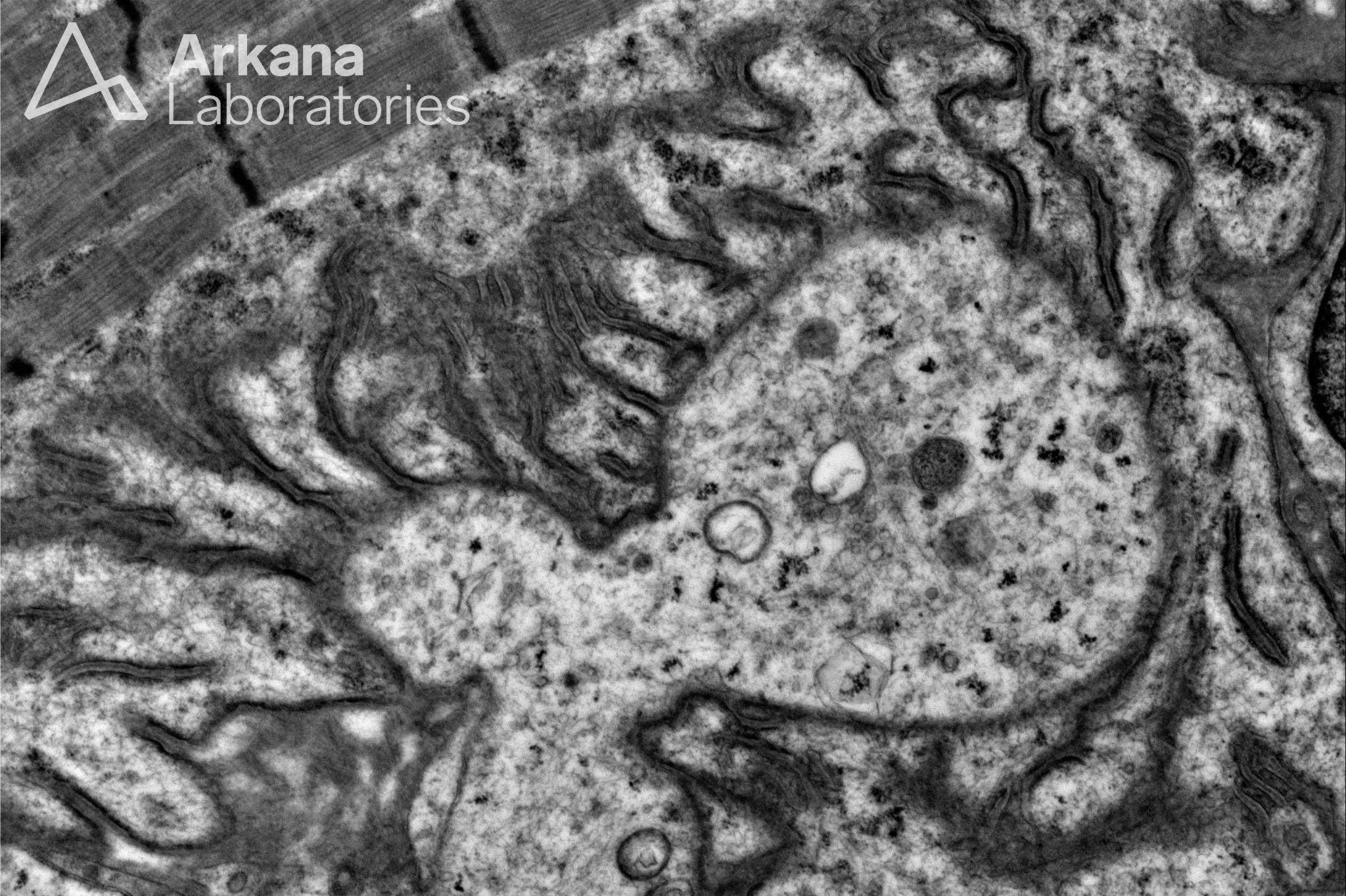
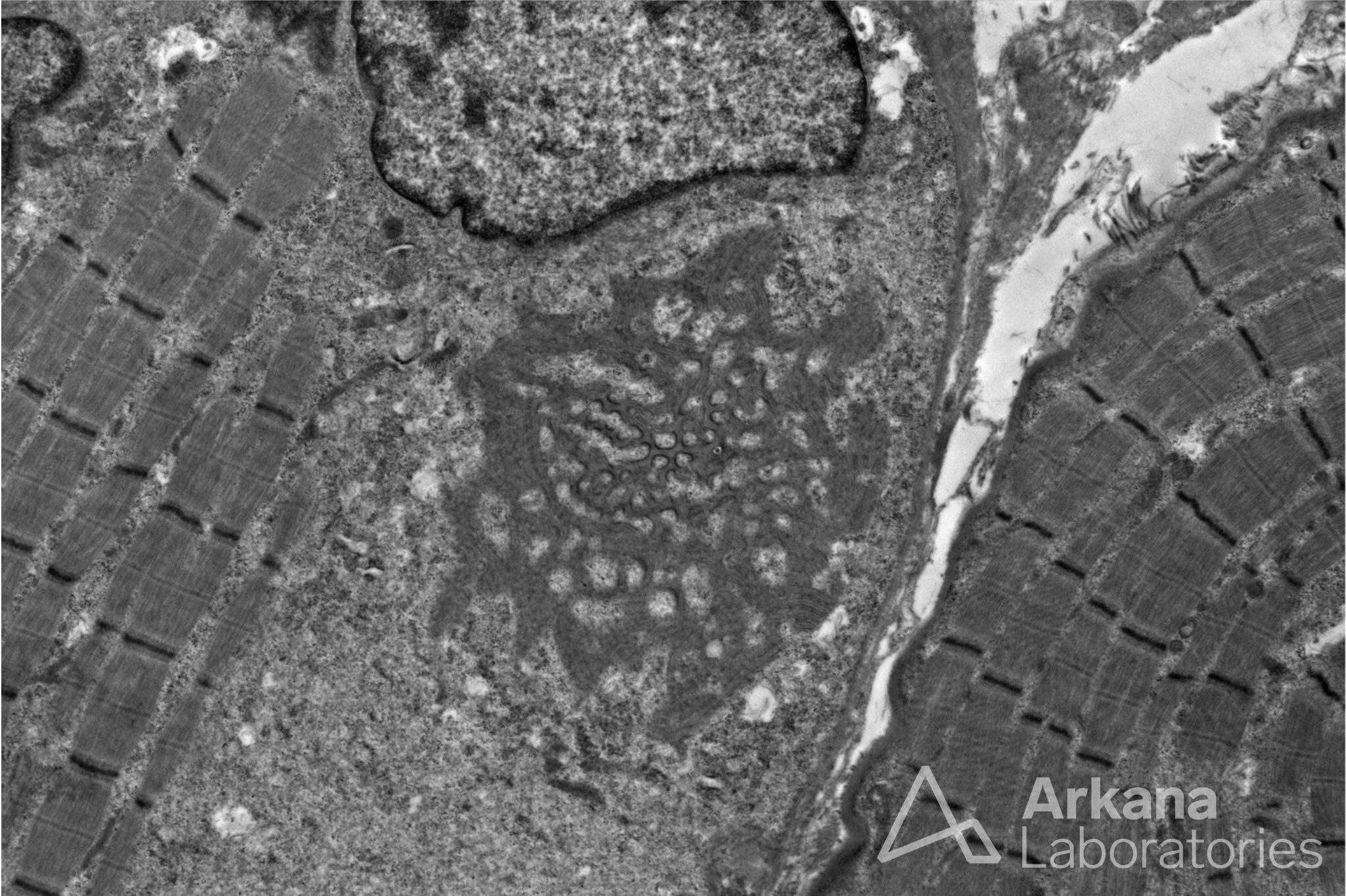
What normal structure is demonstrated in the electron microscopy images?
Answer:
- Neuromuscular junction (aka motor end-plate)
- The first two images appear to be at a branch point of the terminal axon.
- The third image is an enface section of the post-synaptic muscle membrane.
Reference(s) / Additional Reading:
- Nishimune H, Shigemoto K. Practical Anatomy of the Neuromuscular Junction in Health and Disease. Neurol Clin. 2018 May;36(2):231-240. doi: 10.1016/j.ncl.2018.01.009. PMID: 29655446; PMCID: PMC5903580.
- Slater CR. The Structure of Human Neuromuscular Junctions: Some Unanswered Molecular Questions. Int J Mol Sci. 2017 Oct 19;18(10):2183. doi: 10.3390/ijms18102183. PMID: 29048368; PMCID: PMC5666864.
Quick note: This post is to be used for informational purposes only and does not constitute medical or health advice. Each person should consult their own doctor with respect to matters referenced. Arkana Laboratories assumes no liability for actions taken in reliance upon the information contained herein.