This 60-year-old patient presented with muscle weakness and ptosis. Laboratory studies reported normal CPK and negative myositis autoantibody panel. Myasthenia gravis autoantibody panel (AChR, MuSK and LRP4) was negative.
![]()
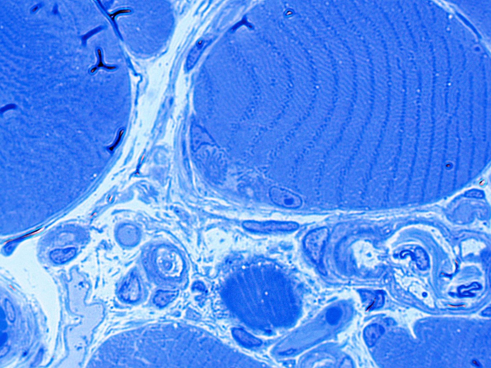
What are the structures indicated by the arrows in Figure #1?
A. Satellite cell
B. Neuromuscular junction
C. Muscle spindle
D. Golgi tendon organ
Answer: Neuromuscular Junction
This high magnification image shows a semilunar area containing two adjacent neuromuscular junctions (black arrows). The oval to round paler areas represent the axon terminal. In this image the motor endplate has an appearance reminiscent of thin flower petals (undulating post-synaptic membrane).
Note the myelinated axons seen in the small nerve twig within the endomysium (white arrow).
Quick note: This post is to be used for informational purposes only and does not constitute medical or health advice. Each person should consult their own doctor with respect to matters referenced. Arkana Laboratories assumes no liability for actions taken in reliance upon the information contained herein.