Muscle biopsy was performed on this 40-year-old patient who presented with proximal lower extremity muscle weakness and normal CPK.
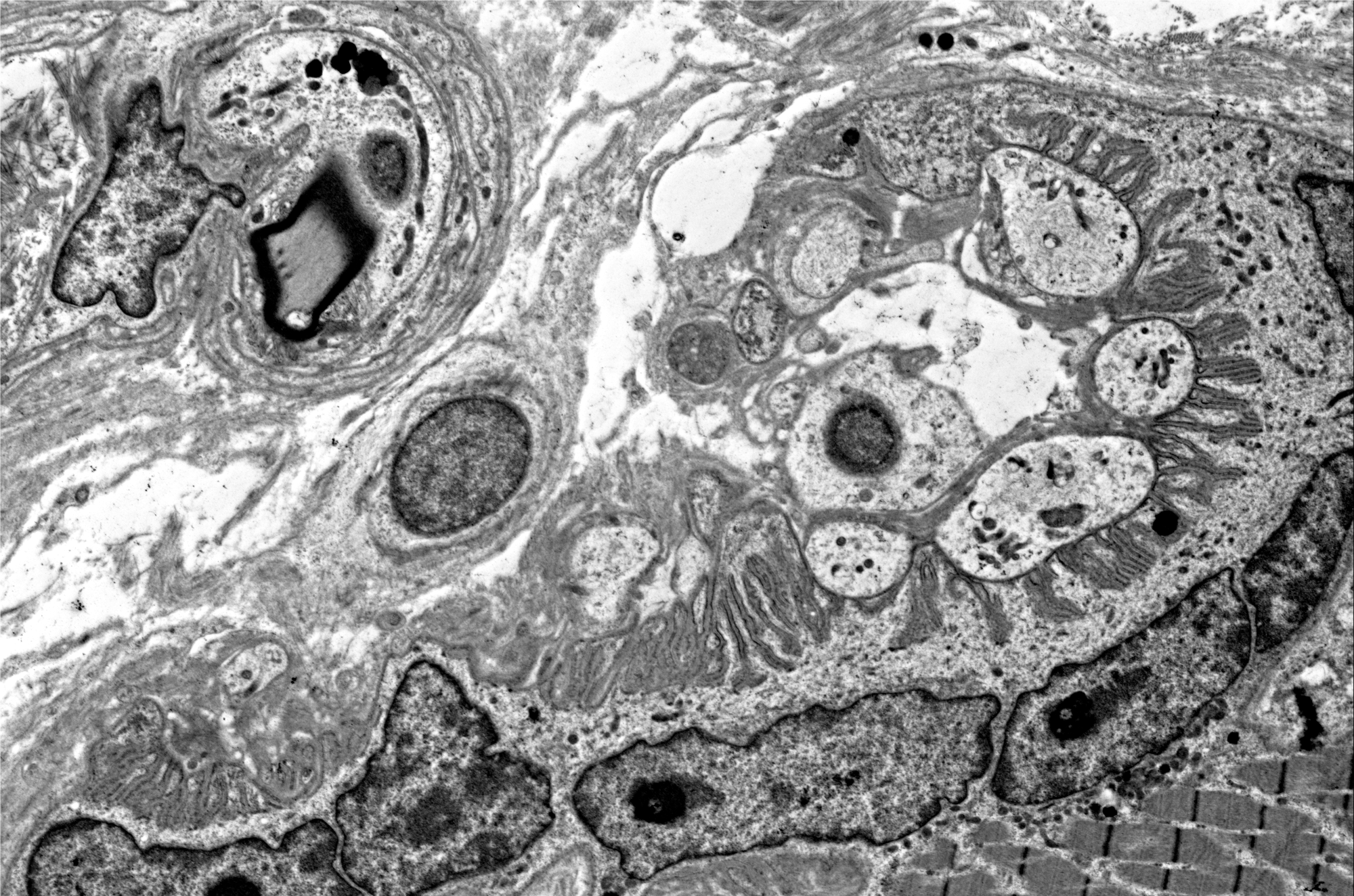
What is the structure demonstrated in Figure #1?
A. Capillary
B. Pacinian corpuscle
C. Neuromuscular junction
D. Parasite
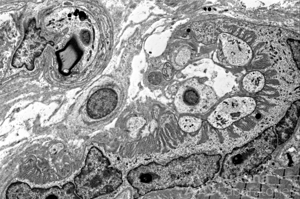
Note the following: A. Nerve twig with myelinated axon B. Terminal branches of motor axon (presynaptic) C. Motor endplate (post-synaptic surface) D. Muscle fiber nuclei *at this magnification it is not possible to appreciate the synaptic cleft (very narrow space between the nerve terminal and motor end plate) or the synaptic vesicles containing neurotransmitter within the terminal nerve endings.
Answer: Neuromuscular Junction
The image shows the components of the neuromuscular junction (AKA myoneural junction). The undulating motor endplate serves to increase the surface area of the post-synaptic membrane.
The region of the motor endplate typically shows associated muscle fiber nuclei and mitochondria.
Reference(s) / additional reading:
https://www.ncbi.nlm.nih.gov/books/NBK470413/
https://www.ncbi.nlm.nih.gov/pmc/articles/PMC5903580/
Braz LP, Ng YS, Gorman GS, Schaefer AM, McFarland R, Taylor RW, Turnbull DM, Whittaker RG. Neuromuscular Junction Abnormalities in Mitochondrial Disease: An Observational Cohort Study. Neurol Clin Pract. 2021 Apr;11(2):97-104. doi: 10.1212/CPJ.0000000000000795. PMID: 33842062; PMCID: PMC8032443.
Quick note: This post is to be used for informational purposes only and does not constitute medical or health advice. Each person should consult their own doctor with respect to matters referenced. Arkana Laboratories assumes no liability for actions taken in reliance upon the information contained herein.