Clinical history
This 60-year-old patient presented with chronic ptosis and mild muscle weakness. CPK levels were within normal limits.
Based on Figures #1 – #4 which of the following is the best diagnosis?
A. Polymyositis
B. IBM
C. Mitochondrial myopathy
D. McArdle disease


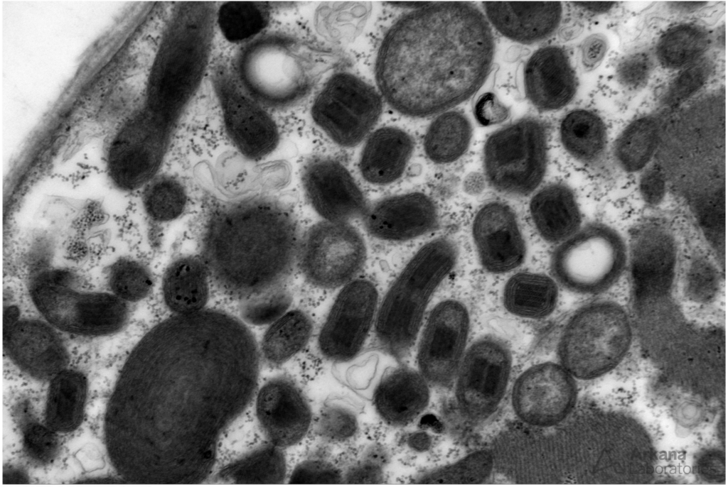
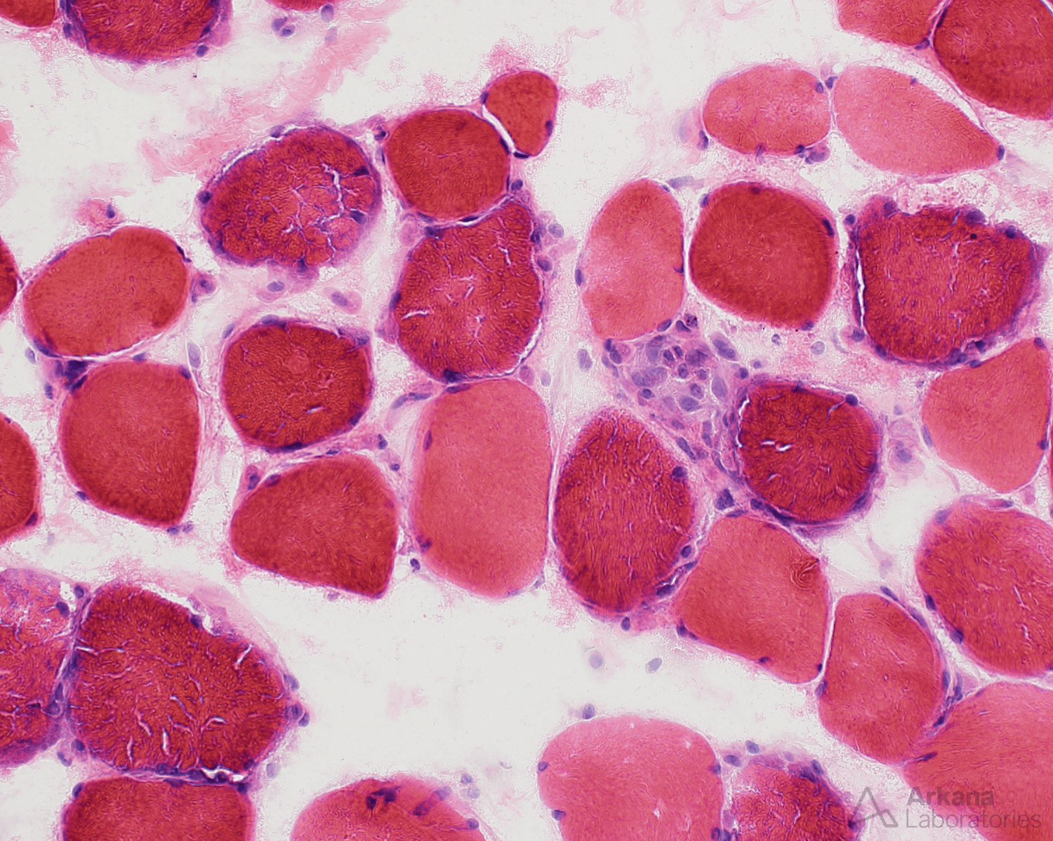
Figure 1: This medium power image shows scattered muscle fibers with mild basophilic tinctoral quality.
Figure 2: A higher power image shows scattered muscle fibers with coarse bright magenta to bluish staining between the sarcomeres and in a subsarcolemmal distribution (ragged-red muscle fibers).
Figure 3: This medium power image shows frequent COX-negative myofibers (blue tinctoral quality with this dual stain preparation)
Figure 4: Electron microscopy shows frequent muscle fibers with mitochondrial abnormalities:
-enlarged
-spirally arranged critae
-paracrystalline inclusions (“parking lot inclusions”)
Correct answer: C. Mitochondrial myopathy
Quick note: This post is to be used for informational purposes only and does not constitute medical or health advice. Each person should consult their own doctor with respect to matters referenced. Arkana Laboratories assumes no liability for actions taken in reliance upon the information contained herein.